
Peptide Research in Tendon and Ligament Biology

Last updated: July 2026
A peptide tendon repair research model is a lab system that tests how small protein chains affect tendon and ligament tissue. Tendons connect muscle to bone. Ligaments connect bone to bone. Both are made mainly of collagen type I in a dense, parallel fiber pattern. This structure gives them tensile strength but limits their blood supply and natural repair speed. According to PubMed (2018), BPC-157 has shown activity in multiple connective tissue repair models including tendon and ligament assays.
Next Level Pharm stocks research-grade peptides for tendon and ligament biology studies. Every vial is HPLC-tested and ships with a certificate of analysis (COA). Average purity across recent batches is 99.4%.
Tendon and ligament repair is a complex process. The poor blood supply to these tissues slows natural repair. Labs use peptides to probe how repair signals can be amplified in low-vascularization tissue models. Understanding the collagen response and tenocyte behavior is a key focus of this research.
Key Takeaways
- Collagen Type I Dominates: Tendons and ligaments are 70-80% collagen type I by dry weight. Research peptides are often studied for their effects on collagen synthesis and cross-linking in fibroblast and tenocyte models.
- BPC-157 in Tendon Models: BPC-157 is a 15-amino-acid peptide studied for promoting angiogenesis and collagen cross-linking in tendon repair assays. It targets both the cell growth and remodeling stages.
- TB-500 and Tenocyte Migration: TB-500 (thymosin beta-4) promotes actin-driven cell migration. Tenocytes, the main cells of tendon tissue, use actin networks to move to injury sites.
- Angiogenesis in Tendon Repair: Tendons have very low blood vessel density. New vessel growth is a key early step in repair. BPC-157 is studied for its ability to promote this step.
- Tenocyte vs. Fibroblast Models: Labs use both tenocyte cell lines and dermal fibroblast cultures to study tendon repair peptides. Each gives different data on collagen production and cell migration.
- COA-Verified Supply: All tendon repair peptides are lyophilized and batch-tested. Lot numbers link to online purity records for each vial.
The sections below expand each point with research context and lab model details.
What Makes Tendons and Ligaments Hard to Study?
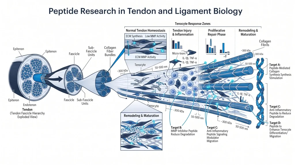
Tendons and ligaments have very limited blood supply. This makes them slow to receive repair signals. The main cells in tendon tissue are tenocytes. These are elongated fibroblast-like cells that sit between collagen fibers. They make and maintain collagen type I. But in dense connective tissue, their movement and signaling are restricted.
When tissue stress occurs, tenocytes must migrate to the site and start repair. They also need growth factor signals from nearby blood vessels. Without good blood supply, these signals arrive slowly. Labs use in vitro scratch assays and collagen gel models to study tenocyte behavior under peptide input. This bypasses the blood supply limit and lets researchers observe cell responses directly.
How Does BPC-157 Work in Tendon Research?
BPC-157 is a 15-amino-acid peptide studied across many connective tissue models. In tendon assays, it is studied for two main effects. First, it promotes angiogenesis. New blood vessel growth brings more repair cells and growth factors to the tendon. Second, it supports collagen cross-linking. Cross-linked collagen fibers form a stronger repair matrix.
BPC-157 also modulates nitric oxide (NO) signaling. NO widens small blood vessels and supports new capillary formation. This is key in tendon tissue where vessel density is naturally low. According to PubMed (2018), BPC-157 showed consistent activity across multiple tendon repair model types. Labs studying tendon biology can browse BPC-157 research vials at Next Level Pharm for COA-verified supply.
What Is TB-500’s Role in Ligament Studies?
TB-500 is thymosin beta-4, a 43-amino-acid peptide. It binds the protein actin, which forms the internal skeleton of cells. Cells use actin to move and change shape. In tendon and ligament tissue, tenocytes and fibroblasts rely on actin networks to migrate to repair sites. TB-500 helps these cells move faster and farther in lab models.
In scratch assays, TB-500 increases the speed of gap closure by tenocyte cell lines. It also reduces swelling markers in stressed connective tissue cultures. According to PubMed (2012), thymosin beta-4 promotes actin-driven cell migration in repair assay systems across multiple tissue types. Browse TB-500 research vials for batch-tested, lyophilized supply.
What Is the Role of Collagen in Tendon Research?
Collagen type I is the main structural protein in tendons and ligaments. It forms long, parallel fibers that run along the axis of mechanical load. These fibers give tendons their tensile strength. In repair, damaged collagen fibers must be replaced. Fibroblasts and tenocytes make new procollagen molecules. These are then assembled and cross-linked into mature fibers.
Cross-linking is a key step. Without good cross-linking, new collagen fibers are weak and prone to failure. Peptides that support cross-linking are studied for their ability to improve repair quality in lab models. GHK-Cu is another compound studied for collagen support. Browse GHK-Cu research vials for a collagen-focused research compound.
How Do Tenocytes Behave in Peptide Assays?
Tenocytes are the main cells of tendon tissue. They are elongated, spindle-shaped cells that sit between collagen fiber bundles. In lab models, tenocytes are isolated and grown in culture. They retain their collagen-producing behavior in culture for several passages. Labs then test peptides on these cultures to see how they change collagen output, migration speed, and gene expression.
Scratch assays test how fast tenocytes close a gap in a culture layer. Collagen gel contraction assays test how strongly tenocytes pull on a surrounding collagen matrix. Both give useful data on how peptides change tenocyte function. BPC-157 and TB-500 are often tested in both assay types to compare their effects on migration and collagen organization.
How Do Tendon Repair Peptides Compare?
Labs choose tendon repair peptides based on which part of the repair process they want to probe.
| Peptide | Primary Target | Mechanism | Key Assay |
| BPC-157 | Angiogenesis, collagen | NO signaling, cross-link support | Tendon repair models, NO assays |
| TB-500 | Tenocyte migration | Actin binding, cell motility | Scratch assay, gel contraction |
| GHK-Cu | Collagen synthesis | Fibroblast activation, MMP action | Cell culture, skin/tendon models |
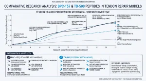
Combining BPC-157 and TB-500 in one study covers both angiogenesis and migration. This gives a broader view of the repair response than either compound alone.
Frequently Asked Questions
What is peptide tendon repair research?
Peptide tendon repair research uses lab models to study how small protein chains affect tendon and ligament biology. Researchers track effects on tenocyte migration, collagen synthesis, angiogenesis, and swelling markers. Common models include tenocyte cell cultures, scratch assays, collagen gel systems, and animal tendon injury models. BPC-157 and TB-500 are the most studied compounds in this area. All research is for lab use only and is not intended for human therapeutic use.
Why are tendons hard to repair in research models?
Tendons have very low blood vessel density compared to other tissues. This limits how fast repair cells and growth factors reach the injury site. Tenocytes, the main repair cells, are slow to migrate in dense collagen matrix environments. These combined factors make tendon repair a slow and complex process. Lab models use peptides to probe how repair signals can be amplified in low-vascularization tissue systems.
How does BPC-157 promote tendon repair in lab models?
BPC-157 promotes angiogenesis and collagen cross-linking in tendon repair assays. It modulates nitric oxide (NO) signaling to widen small blood vessels and support new capillary growth. Better blood supply brings more repair cells to the site. BPC-157 also supports collagen cross-linking, which strengthens the repair matrix. According to PubMed (2018), BPC-157 showed consistent activity across multiple tendon repair model types in preclinical studies.
What does TB-500 do in tenocyte migration assays?
TB-500 binds actin, the protein that forms the internal skeleton of cells. By modulating actin dynamics, TB-500 helps tenocytes migrate faster to repair sites in scratch assay models. According to PubMed (2012), thymosin beta-4 promotes actin-driven cell migration in repair assay systems. TB-500 also reduces swelling markers in stressed connective tissue cultures, making it useful for studying the acute repair phase in tendon models.
What is collagen type I and why does it matter in tendon research?
Collagen type I is the main structural protein in tendons and ligaments. It forms long, parallel fibers that run along the axis of mechanical load. These fibers give tendons their tensile strength. In repair, damaged fibers must be replaced by new ones made by tenocytes and fibroblasts. Cross-linking of new fibers is a key quality step. Peptides that support collagen synthesis and cross-linking are studied for their ability to improve repair matrix quality in lab models.
What are tenocytes and how are they studied in peptide research?
Tenocytes are elongated, spindle-shaped cells that sit between collagen fiber bundles in tendon tissue. They make and maintain collagen type I. In lab studies, tenocytes are isolated and grown in culture. Researchers test peptides on these cultures to measure changes in collagen output, migration speed, and gene expression. Scratch assays measure migration speed. Collagen gel contraction assays measure how strongly tenocytes pull on a surrounding collagen matrix.
Why is angiogenesis a key step in tendon repair?
Angiogenesis is the growth of new blood vessels. Without new vessels, a tendon repair site cannot receive repair cells, growth factors, and oxygen in adequate amounts. Tendon tissue already has low native vessel density, which makes this step even more important. BPC-157 is studied for its angiogenic activity in tendon models. Labs track new vessel density and tube formation as endpoints for angiogenesis in tendon repair assays.
How are scratch assays used in tendon biology research?
A scratch assay creates a gap in a layer of cells grown on a dish. Researchers then measure how fast cells close the gap. This models cell migration in tissue repair. In tendon research, scratch assays use tenocyte cell lines to test how peptides change gap-closure speed. TB-500 and BPC-157 are both tested in scratch assays. Results show how each peptide changes the speed and pattern of tenocyte migration under controlled lab conditions.
Can BPC-157 and TB-500 be studied together in one assay?
Yes, labs often use both compounds in a combined study design. BPC-157 covers angiogenesis and collagen cross-linking. TB-500 covers actin-driven cell migration. Together, they target two different phases of the tendon repair process. Multi-compound assays allow researchers to observe how angiogenic and migratory signals interact in the same model. All compounds must be COA-verified and HPLC-tested to ensure purity and lot consistency across a combined study run.
Summary
Tendon and ligament peptide research studies how small protein chains affect connective tissue repair in lab models. BPC-157 targets angiogenesis and collagen cross-linking in low-vascularization tissue. TB-500 promotes tenocyte migration via actin dynamics. GHK-Cu supports collagen synthesis and matrix remodeling in fibroblast cultures.
Labs use scratch assays, collagen gel models, and animal tendon models to gather data. Each compound targets a different part of the repair cascade. Multi-compound designs cover more of the repair process in a single study.
What Should You Do Next?
Researchers should identify the tendon or ligament repair stage under study. Select a peptide and assay endpoint that match the target mechanism. Confirm HPLC purity, mass spec identity, and the lot number on the COA before each run. Researchers sourcing these lab peptides can shop research peptides with full COA and lot traceability.
People Also Read
- BPC-157 Research Peptide: Lab-Grade Vials for Tissue Research
- TB-500 Research Peptide: Thymosin Beta-4 for Lab Use
About the Author
Next Level Pharm Research Team
Alex M covers peer-reviewed findings in peptide science for Next Level Pharm, a US-based supplier of research-grade peptides verified to ≥99% purity via HPLC and mass spectrometry on every batch.
Disclaimer: For research purposes only. Not intended for human consumption. Next Level Pharm products are not intended for diagnostic, therapeutic, or medicinal use. This content does not constitute medical advice. Always consult a licensed healthcare professional before making any health-related decisions.