
IGF-1 LR3 vs MGF: Muscle Research Compared

Last updated: July 2026
An IGF-1 LR3 vs MGF research comparison is a review of two peptides produced from the insulin-like growth factor-1 (IGF-1) gene. Both are studied for distinct roles in muscle tissue research. IGF-1 LR3 has a half-life of 20-30 hours in lab systems. MGF clears within minutes. These differences make them useful tools for studying growth-factor signaling in the lab.
Next Level Pharm supplies these research peptides for lab analysis. It holds an average purity of 99.4% across the last 100 batches. Each vial is tested by HPLC and mass spec. Samples are lyophilized (freeze-dried) for room-temperature stability. No cold chain is needed during shipping.
This comparison covers how small structural changes alter the action of growth-signaling molecules. By comparing these peptides, researchers assess how they trigger different tissue responses in lab models. This helps clarify how peptide sequence changes shape cellular signaling.
Key Takeaways
- Extended Stability: IGF-1 LR3 has a 20-30 hour half-life. Structural changes block IGF-binding protein (IGFBP) binding, which extends its time in lab systems.
- Local Signaling: MGF acts at the site of stress. Its half-life is just minutes, making it a local regulator.
- Shared Receptor: Both peptides bind to the IGF-1 receptor (IGF-1R), though their binding profiles differ.
- Research Roles: IGF-1 LR3 is studied for systemic cell growth. MGF is studied for satellite cell activation and local tissue repair.
- Gene Origin: Both come from the IGF-1 gene. They differ in how they are made and where they act.
- Protocol Design: Each compound needs distinct timing in research protocols due to their half-life differences.
Researchers on muscle tissue models choose between these peptides based on the scope of their study. IGF-1 LR3 suits tests focused on systemic, long-duration signaling. MGF suits protocols built to capture acute, local responses to mechanical stress. Knowing the structural basis for these differences is key when designing comparative studies.
What Is Insulin-Like Growth Factor-1 Long R3?
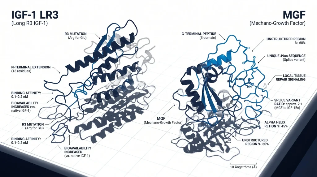
Insulin-like growth factor-1 Long R3 (IGF-1 LR3) is a synthetic analog of human IGF-1. It is built to boost the potency and half-life of the native molecule. This peptide has an N-terminal extension and one point mutation. These changes limit its contact with control proteins. This lets the molecule avoid the fast metabolic clearance that typically affects native factors in lab settings.
The Long R3 change involves two key edits to the base sequence. First, arginine replaces glutamic acid at the third position. Second, it adds a 13-amino acid extension to the N-terminus. According to NCBI (1992), these changes must reduce the affinity of the molecule for IGF-binding proteins. Lower binding means the analog stays active longer, with a half-life over 20 hours.
The key changes in IGF-1 LR3 are listed below.
- Arginine substitution: replaces glutamic acid at the third position of the sequence.
- N-terminal extension: adds 13 amino acids to the base sequence.
- IGFBP avoidance: lowers binding for circulating control proteins.
- Extended half-life: 20-30 hours versus a few hours for native IGF-1.
Next Level Pharm stocks IGF-1 LR3 as a COA-verified research peptide. Every lot is tested by HPLC and mass spec. Browse the Fitness and GH peptide category to view available vials.
What Is Mechano Growth Factor (MGF)?
Mechano Growth Factor (MGF) is a splice variant of the IGF-1 gene. It expresses in muscle tissue during periods of mechanical stress. This variant comes from the IGF-1 gene but goes through distinct RNA splicing when muscle fibers face tension or injury. Research shows this process serves as a local signaling action for muscle upkeep.
The E-domain is a unique C-terminal peptide segment that marks MGF. This region lets the molecule act apart from systemic liver-derived IGF-1. When muscle satellite cells detect this signal, they activate to help repair damaged fibers. This activity is studied for muscle fiber growth in lab models.
Stem cell activation can lead to more myonuclei (muscle cell nuclei). This gives a structural base for fiber adaptation. For related reading, see PEG-MGF research and myocyte studies.
How Do Their Mechanisms of Action Differ in Research?
IGF-1 LR3 research focuses on its systemic effects. It promotes cell growth through hyperplasia by binding to receptors across the body. MGF research centers on its local autocrine and paracrine signaling to start site-specific muscle repair. These two paths are distinct, making them useful for comparative studies.
The key difference is their stability and reach. IGF-1 LR3 is modified to block binding to plasma proteins. This gives it a long half-life and lets it move through the body. MGF is a splice variant made at the site of damage to act on nearby cells. This local action keeps repair signals where they are needed.
Each compound gives unique data on how distinct paths link the response to physical load. For background on how half-life shapes research design, see what is peptide half-life and stability in research.
What Does Research Show on Their Receptor Interactions?
Both peptides mainly interact with the IGF-1 receptor (IGF-1R), but their binding profiles differ much. Research shows their molecular structures dictate how they engage target cells in assay protocols. IGF-1 LR3 is built for high receptor affinity while avoiding the blocking effect of IGF-binding proteins. MGF acts through the IGF-1R but uses its unique E-domain to trigger specific cell responses.
IGF-1 LR3 avoids capture by circulating IGF-binding proteins. This lets it hold steady activity at the receptor site for longer. According to research at NCBI (2002), the unique E-domain of MGF is key to its ability to activate satellite cells. Binding differences serve as a main variable for telling apart systemic signaling from local fiber repair in lab models.
Which Is Studied for Muscle Cell Hyperplasia vs Repair?
IGF-1 LR3 is mainly studied for its effect on muscle cell hyperplasia. MGF is studied for its role in local tissue repair. Research shows that IGF-1 LR3 promotes systemic growth by driving the division of cells to create new muscle fibers. MGF acts as a mechanical signaling molecule to keep tissue intact after physical stress.
These distinctions guide research protocol selection.
- IGF-1 LR3: systemic cell growth, multi-tissue studies, long study windows.
- MGF: local repair, satellite cell protocols, acute post-stress studies.
The main focus of MGF research is the activation of satellite cells in response to damaged tissue. These cells move to the site of stress and fuse with fibers, adding to local growth. Next Level Pharm makes both peptides as COA-verified research compounds with lot-specific analysis docs.
How Do Their Half-Lives Affect Research Protocols?
The large gap in half-life shapes study design. IGF-1 LR3 has a 20-30 hour half-life. This allows for fewer delivery cycles while holding stable systemic levels during the study. This duration is useful for tracking long-term changes in cell growth across tissue types.
The minutes-long half-life of MGF needs a focused approach in lab settings. Researchers must plan for fast delivery near the target site to capture its acute effects. Because this molecule clears fast, protocols often involve delivery right after inducing mechanical stress on muscle fibers. This design keeps the peptide active during the key window for satellite cell activation and repair.
How Do IGF-1 LR3 and MGF Compare?
| Feature | IGF-1 LR3 | MGF |
| Peptide type | Synthetic analog | Splice variant |
| Source | Engineered (modified IGF-1) | Alternative IGF-1 gene splicing |
| Half-life | 20-30 hours | Minutes |
| Site of action | Systemic (whole body) | Local (muscle tissue) |
| Primary receptor | IGF-1R | IGF-1R |
| Key research use | Cell hyperplasia, systemic signaling | Satellite cell activation, local repair |
Frequently Asked Questions
What is the difference between IGF-1 LR3 and MGF?
IGF-1 LR3 is a synthetic analog built for extended time in lab systems through structural changes. It acts mainly in systemic paths to drive cell growth. MGF is a locally expressed splice variant of the IGF-1 gene. Muscle tissue makes it in response to mechanical stress. Scientists tell them apart by their kinetic profiles and their roles in either systemic signaling or local tissue repair.
Which peptide is studied for muscle cell research, IGF-1 LR3 or MGF?
Selection depends on the specific research question. IGF-1 LR3 is used when researchers need to study cell hyperplasia or systemic growth signals over a 24-hour period. MGF is used for research on acute, local tissue response after mechanical stress. No objective superiority exists because each compound probes a unique biological path. Researchers choose the peptide that fits the scope of their study.
Can IGF-1 LR3 and MGF be alternated in a research protocol?
Protocols can include both peptides if the test aims to observe how different paths contribute to tissue control. Because IGF-1 LR3 has a long half-life and MGF has a rapid one, researchers must time their observations with care. Using both peptides lets scientists track long-term cell activity alongside acute response events. Studies must account for the activation windows of both compounds to keep data readable.
What gives IGF-1 LR3 its long half-life?
The long half-life of IGF-1 LR3 comes from two key chemical changes to the native protein. Researchers replaced glutamic acid with arginine at the third position of the sequence. They also added a 13-amino acid extension to the N-terminus. According to NCBI (1992), these changes block binding to IGF-binding proteins. This lowers the rate of metabolic clearance and extends the active life of the molecule.
How is MGF produced in response to muscle stress?
Muscle tissue makes MGF naturally when structural stress or damage occurs. This involves alternative splicing of the IGF-1 gene in response to mechanical load. According to NCBI (2002), this variant surges during the early stages of muscle adaptation. This local signal keeps the peptide at the exact site of stress. It does not travel through the whole body during this time.
Do IGF-1 LR3 and MGF bind to the same receptor?
Both peptides use the IGF-1 receptor (IGF-1R) to start their primary signaling cascades. But their binding patterns differ due to the unique structures of each molecule. IGF-1 LR3 reaches receptors through systemic spread, signaling for cell growth across many tissues. MGF binds to local receptors to trigger repair within specific muscle fibers. The receptor target is the same, but the outcome of binding varies by molecule.
Is MGF research focused on local or systemic effects?
Research on MGF is almost entirely focused on its local, autocrine, and paracrine effects within muscle environments. Its half-life is very brief. So it does not act as a systemic hormone like native IGF-1 or its synthetic analogs. Researchers study MGF to see how muscles manage their own repair without relying on global hormonal signals. Peer-reviewed data shows that MGF remains a local regulator of tissue integrity.
What is the significance of the MGF E-domain?
The E-domain is a distinct tail sequence found at the C-terminus of the MGF molecule. This region sets MGF apart from other IGF-1 isoforms and is vital for its activity in repair scenarios. According to NCBI (2004), the E-domain regulates receptor-specific interactions that set MGF apart from systemic growth factors. This structural feature lets MGF start the activation of satellite cells, supporting local repair of fibers.
What are muscle satellite cells and what is MGF’s role in their activation?
Muscle satellite cells are stem cells located between the basement membrane and the plasma membrane of muscle fibers. They stay inactive until a repair signal triggers them to re-enter the cell cycle. MGF acts as a mitogen (a substance that drives cell division). It prompts satellite cells to grow and fuse with existing fibers. Research shows this activation is a key step for keeping muscle structure after physical damage or intense use.
How does IGF-1 LR3 compare to native IGF-1 in research settings?
Native IGF-1 is made mainly in the liver and moves through the body. But it binds readily to IGF-binding proteins (IGFBPs), which limits how much stays active. IGF-1 LR3 is built to bypass this limit. Its structural changes much reduce IGFBP binding. This lets the analog hold greater receptor availability during the study window. Researchers use this to study how prolonged IGF-1R activation differs from the pulsatile activity of the native hormone.
Summary
IGF-1 LR3 and MGF both come from the IGF-1 gene, but they serve distinct research purposes. IGF-1 LR3 is a synthetic analog with a 20-30 hour half-life. It is studied for systemic cell growth and broad anabolic signaling across tissue types. MGF is a natural splice variant with a half-life of just minutes. It is studied for local satellite cell activation and tissue repair.
Both peptides target the IGF-1 receptor (IGF-1R), but their outcomes differ based on structural design. IGF-1 LR3 avoids IGFBP capture to hold systemic activity. MGF keeps its activity at the site of mechanical stress. These differences make them useful tools for researchers studying the control of muscle tissue biology.
What Should You Do Next?
Researchers should match IGF-1 LR3 or MGF to the pathway and timing needs of the planned model. Compare sequence, stability, and half-life data before selecting a compound. Review the lot COA and record HPLC purity, mass spec identity, and the lot number in the protocol. Researchers sourcing these lab peptides can shop research peptides with full COA and lot traceability.
People Also Read
- IGF-1 LR3 Research: What Studies Show About This Growth Factor Analog
- MGF Research: Mechano Growth Factor Mechanism and Study Findings
About the Author
Next Level Pharm Research Team
Alex M covers peer-reviewed findings in peptide science for Next Level Pharm, a US-based supplier of research-grade peptides verified to ≥99% purity via HPLC and mass spectrometry on every batch.
Disclaimer: For research purposes only. Not intended for human consumption. Next Level Pharm products are not intended for diagnostic, therapeutic, or medicinal use. This content does not constitute medical advice. Always consult a licensed healthcare professional before making any health-related decisions.