IGF-1 Signaling: PI3K/Akt/mTOR Pathway Research

Last updated: July 2026
An IGF-1 signaling pathway research model is a lab system used to map the cellular network between receptor binding and gene output. It links surface receptor binding to gene expression, protein production, and cell survival. IGF-1, or insulin-like growth factor 1, binds to its receptor (IGF-1R) at the cell membrane. This binding starts a cascade of chemical signals that govern how cells grow and divide. According to NCBI (2018), this pathway is key for normal tissue growth and repair in lab models.
Next Level Pharm supplies COA-verified, research-grade peptides linked to the IGF-1 axis. Each vial is tested to 99.4% average purity across the last 100 batches. Verification uses HPLC (high-performance liquid chromatography) and mass spectrometry on every lot. Peptides ship lyophilized, or freeze-dried, with an average 48-hour dispatch across the USA.
The IGF-1 cascade splits into two major branches at the receptor level. Each branch targets different cell functions. Understanding both branches helps researchers map how a single growth signal can produce many different cell outcomes.
Key Takeaways
- IGF-1 Receptor activity: IGF-1 binds to its surface receptor, IGF-1R, and starts activation events. These events open two major signaling branches.
- PI3K/Akt Branch: Activated PI3K converts PIP2 to PIP3. This recruits Akt to the membrane and turns on growth signals.
- mTOR as Anabolic Switch: Akt blocks the TSC1/TSC2 complex and frees Rheb. This lets mTORC1 drive protein production.
- Cell Survival Control: Akt blocks pro-cell-death proteins Bad and FOXO. This keeps cells from triggering programmed death signals.
- MAPK/ERK Branch: The Ras-Raf-MEK-ERK cascade runs in parallel. It governs gene expression and cell division timing.
- Disease Model Research: Scientists study IGF-1 pathway dysfunction in cancer and metabolic models. Overexpression leads to uncontrolled cell growth.
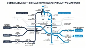
These two branches work in parallel but target different cell functions. PI3K/Akt/mTOR drives energy, survival, and protein output. MAPK/ERK controls gene expression and when the cell divides. Both are active areas of study in lab models.
What Is the Core IGF-1 Signaling Cascade?
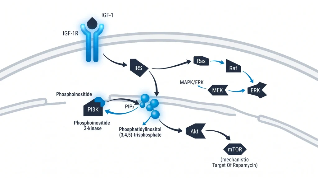
IGF-1 binds IGF-1R and triggers receptor self-activation. This creates docking sites for IRS (insulin receptor substrate) proteins. IRS proteins bridge the receptor to two branches: PI3K/Akt/mTOR and Ras/MAPK. These branches then connect surface events to cell responses. According to NCBI (2018), these nodes adjust internal cell states in response to external signals.
PI3K/Akt mainly controls protein production, energy balance, and cell growth. MAPK controls cell division and gene transcription. Both branches alter the activity of gene switches, which changes which genes turn on. The PI3K and MAPK branches can also cross-talk, sharing signals at certain nodes.
How Does the PI3K/Akt Pathway Get Activated?
IGF-1 binds IGF-1R and triggers self-activation. This creates docking sites on the receptor tail. IRS proteins bind these sites and get activated. They then recruit PI3K (phosphoinositide 3-kinase) to the membrane. PI3K converts PIP2 to PIP3. PIP3 is a lipid messenger that docks Akt at the membrane. Once docked, Akt is activated and turns on. Per PubMed (2019), this chain of events starts downstream survival and metabolic signals.
IRS proteins act as scaffolds that hold multiple signaling proteins close together. This speeds up signal transfer inside the cell. Akt then targets many proteins that block cell death, boost metabolism, and promote growth. Active Akt phosphorylates Bad, a protein that would otherwise trigger cell death. It also controls FOXO gene switches, which regulate survival genes.
What Does mTOR Do in This Pathway?
Akt phosphorylates and blocks the TSC1/TSC2 complex. TSC1/TSC2 normally acts as a brake on a small protein called Rheb. Once blocked, Rheb becomes free and active. Active Rheb turns on mTORC1 (mechanistic target of rapamycin complex 1). mTORC1 then targets S6K1 and 4E-BP1 to boost protein production. Per Journal of Peptide Science (2022), mTORC1 drives an anabolic cell state in lab models.
When mTORC1 is active, cells shift into a growth and building mode. S6K1 and 4E-BP1 both affect how ribosomes translate mRNA into proteins. Blocking mTORC1 with rapamycin reverses this state in lab models. Next Level Pharm provides peptides such as IGF-1 LR3 for researchers who study how upstream IGF-1 signals reach mTOR. Energy and nutrient levels also feed into mTORC1, making it a central node.
How Does IGF-1 Signaling Control Cell Survival?
Akt blocks cell death, or programmed cell death, by targeting Bad and FOXO gene switches. When Akt phosphorylates Bad, Bad cannot enter the mitochondrial membrane. This stops Bad from triggering cell death signals. When Akt phosphorylates FOXO proteins, they cannot enter the nucleus or activate death-related genes. This keeps cells alive and functioning. According to PubMed (2019), Akt is a major survival switch in lab cell models.
FOXO proteins belong to a family of gene switches that control stress response genes. When IGF-1 is present and Akt is active, FOXO stays out of the nucleus. Bad is also a pro-cell-death marker: its activation by Akt neutralizes its cell-death function. Researchers use IGF-1 DES, another COA-verified research peptide, to study how structural analogs affect this survival branch in vitro.
How Does the MAPK/ERK Branch Work?
IGF-1R also activates the Ras-Raf-MEK-ERK branch in parallel with PI3K/Akt. ERK (outside the cell signal-regulated kinase) moves into the nucleus once active. There it acts on gene switches c-Fos and c-Jun. These factors control the timing of the cell cycle. According to PubMed (2023), the MAPK branch links growth signals to gene expression in lab models.
MAPK/ERK focuses on gene expression and when the cell divides. PI3K/Akt focuses on energy and survival. Both branches can reinforce each other under strong IGF-1 signals. Below is a summary of how they differ:
| Feature | PI3K/Akt/mTOR | MAPK/ERK |
| Primary role | Cell survival and protein production | Gene expression and cell division |
| Key effector | mTORC1 | ERK and gene switches |
| Output | Anabolic growth state | Cell cycle progression |
Why Is This Pathway Studied in Disease Models?
Overexpressed or mutated IGF-1 pathway components appear in many cancer types. These mutations let cells bypass normal growth controls. They also help cells resist programmed death. Metabolic models also use this pathway to study insulin resistance and glucose signaling overlap. Per PubMed (2023), researchers study IGF-1 pathway gain-of-function in multiple disease contexts.
Common lab study contexts include:
- Oncology models: studying uncontrolled cell growth linked to IGF-1R overexpression.
- Metabolic models: examining insulin resistance and cross-talk with glucose signaling.
- Aging research: observing how pathway activity changes affect cell longevity in vitro.
Researchers studying the IGF-1 axis can browse IGF-1 LR3 and related peptides Every vial ships with a COA and lot number.
Also see: MGF Research: Mechano Growth Factor Mechanism and Study Findings for a related GH-axis peptide that activates its own downstream signaling cascade.
Frequently Asked Questions
What is the IGF-1 signaling pathway?
The IGF-1 signaling pathway is a cell communication network. It starts when IGF-1, or insulin-like growth factor 1, binds to its receptor at the cell surface. This binding event triggers two major internal branches: PI3K/Akt/mTOR and Ras/MAPK/ERK. According to NCBI (2018), these nodes control cell growth, survival, and protein production in lab models. The pathway is a core subject in cell biology and disease model research.
How does IGF-1 activate PI3K and Akt?
IGF-1 binds IGF-1R and triggers self-activation. This creates docking sites for IRS proteins, or insulin receptor substrate proteins. IRS proteins then recruit PI3K. PI3K converts PIP2 to PIP3, a lipid messenger that pulls Akt to the membrane. Once at the membrane, Akt gets activated and activates. According to PubMed (2019), this chain of events turns on downstream survival and growth signals in lab models.
What role does IGF-1R play in cell growth research?
IGF-1R, or the IGF-1 receptor, sits at the cell membrane and acts as a gatekeeper. It receives the external IGF-1 signal and translates it into internal instructions. When IGF-1 binds, the receptor undergoes a shape change that starts activation. This single binding event sets off two separate internal branches. Researchers study IGF-1R to understand how cells read growth signals and convert them into changes in gene activity and protein output.
What is the difference between IGF-1 and insulin signaling?
IGF-1 and insulin share structural similarities and both activate PI3K/Akt. But their outputs differ. IGF-1 signaling focuses on cell growth, protein production, and survival. Insulin signaling mainly controls glucose transport into cells. The two pathways overlap at shared nodes but diverge at downstream targets. According to NCBI (2018), researchers study this overlap to understand how metabolic and growth signals cross-talk in lab tissue models.
How are IGF signaling pathways regulated?
Several control systems keep the IGF-1 pathway from staying active too long. IGFBPs, or IGF binding proteins, sequester IGF-1 in the blood and limit how much reaches cell receptors. Inside the cell, mTOR sends negative feedback that reduces upstream activity. PTEN, a phosphatase enzyme, converts PIP3 back to PIP2 to slow Akt. According to PubMed (2023), these control layers keep cell growth in check in normal lab models.
What are IGF binding proteins?
IGF binding proteins, or IGFBPs, are a family of six proteins that bind IGF-1 in blood and tissue. They control how much free IGF-1 is available to reach cell surface receptors. By holding IGF-1 in reserve, IGFBPs shape the timing and strength of receptor signals. Some IGFBPs also have direct effects on cells. Researchers use IGFBPs in lab models to fine-tune IGF-1 availability and study how signal duration affects cell behavior and gene expression.
What is mTORC1’s relationship to IGF-1?
mTORC1 is the final growth-control switch in the PI3K/Akt branch. Akt releases the brake on mTORC1 by phosphorylating and blocking the TSC1/TSC2 complex. This frees a small protein called Rheb. Active Rheb turns on mTORC1. Once on, mTORC1 targets S6K1 and 4E-BP1 to drive protein production. According to the Journal of Peptide Science (2022), mTORC1 links IGF-1 receptor activity to anabolic cell states in lab models.
How does MAPK/ERK differ from PI3K/Akt?
PI3K/Akt controls cell survival, energy balance, and protein output. MAPK/ERK controls gene expression and cell cycle timing. Both branches start at IGF-1R but diverge quickly. ERK moves into the nucleus and acts on gene switches like c-Fos and c-Jun. These factors adjust which genes turn on. According to PubMed (2023), the two branches run in parallel and can reinforce or balance each other based on signal strength.
What research peptides are linked to the IGF-1 axis?
IGF-1 LR3 and IGF-1 DES are two modified analogs used in IGF-1 axis research. IGF-1 LR3 has an extended half-life because it binds poorly to IGFBPs. IGF-1 DES is a truncated form with higher receptor selectivity at the cell surface. Both are COA-verified, research-grade peptides. Researchers use them in lab models to study how changes in IGF-1 structure affect downstream signaling through PI3K, Akt, and mTOR.
How do PI3K/Akt/mTOR blockers work in research models?
blockers target specific kinase enzymes or block docking sites at the cell membrane. A PI3K blocker stops PIP2-to-PIP3 conversion. An Akt blocker blocks Akt activation directly. An mTOR blocker, like rapamycin, binds an mTOR regulatory protein and pauses downstream targets. Researchers use these tools in lab models to map which nodes are needed for cell growth, survival, and response to external signals.
Summary
The IGF-1 signaling pathway links a surface binding event to deep cell changes. IGF-1 binds IGF-1R and starts IRS docking. IRS proteins feed two branches: PI3K/Akt/mTOR and Ras/MAPK/ERK. PI3K/Akt drives protein production and cell survival. MAPK/ERK drives gene expression and cell division. Together, these branches let one external signal shape many internal cell outcomes.
mTORC1 sits at the center of the anabolic branch. Akt releases its brake, and mTORC1 turns on protein synthesis machinery. FOXO and Bad keep survival signals in balance. All of this plays out in lab models, not in living subjects.
Next Level Pharm supplies batch-tested, COA-verified research peptides linked to the IGF-1 axis. Every lot is tested by HPLC and mass spec before dispatch.
What Should You Do Next?
Researchers should map the PI3K, Akt, and mTOR nodes that matter to the planned IGF-1 model. Select suitable blockers and controls from the cited protocols. Confirm the analog identity, HPLC purity, and lot number on the COA before any in vitro work. Researchers sourcing these lab peptides can shop research peptides with full COA and lot traceability.
People Also Read
- IGF-1 LR3 Research: What Studies Show About This Growth Factor Analog.
- Humanin Research: Mitochondria-Derived Peptide and Cell Signaling Studies.
About the Author
Next Level Pharm Research Team
Alex M covers peer-reviewed findings in peptide science for Next Level Pharm, a US-based supplier of research-grade peptides verified to ≥99% purity via HPLC and mass spectrometry on every batch.
Disclaimer: For research purposes only. Not intended for human consumption. Next Level Pharm products are not intended for diagnostic, therapeutic, or medicinal use. This content does not constitute medical advice. Always consult a licensed healthcare professional before making any health-related decisions.