Humanin Research: Mitochondria-Derived Peptide and Cell Signaling Studies

Last updated: May 2026
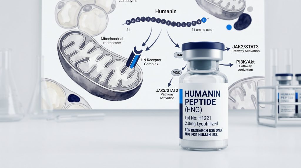
A humanin peptide is a 21-amino acid compound encoded in mitochondrial DNA. Mitochondria are the energy-making parts inside cells. Humanin was first described in 2001 from a study of neural cell survival. It binds to receptors on cell surfaces and starts signal chains that change cell survival and stress responses. Researchers study humanin in neural, heart, and cell models to learn how mitochondria send signals outside the cell. All data here comes from lab and animal models only.
Next Level Pharm supplies research-grade humanin peptide verified to at least 99% purity by HPLC and mass spec. Each lot ships as a freeze-dried solid with a lot-specific COA. Vials are stable during transit at room temperature from US-based stock.
The parts below cover the origin of humanin, its cell signal steps, and findings from published neural and heart cell studies.
Key Takeaways
- Origin: Humanin is encoded in the 16S rRNA gene inside mitochondrial DNA. It is a 21-amino acid peptide.
- Receptors: Humanin binds to cell surface receptors CNTFR, WSX-1, and gp130, which form a three-part complex.
- Signaling: Receptor binding starts the JAK2/STAT3 and PI3K/Akt pathways. Both chains affect cell death and stress signals.
- Neural Studies: Lab studies show humanin cuts apoptosis markers in neural cell lines exposed to amyloid-beta.
- Heart Cell Studies: Humanin has been studied in heart cell models for its effect on oxidative stress and energy output markers.
- Supply Standard: Research needs a lot-specific COA with HPLC purity at or above 99% and mass spec check.
The parts below expand on each point with published study findings.
What Is the Origin of Humanin Peptide?
Humanin is encoded in the 16S rRNA gene inside mitochondrial DNA. Most proteins made by mitochondria stay inside the cell part. Humanin is one of a small group that gets sent out and acts outside the cell. This makes it a mitochondria-derived peptide, or MDP.
The full humanin sequence is 21 amino acids. It is stable as a synthetic peptide at the same length as the natural form. According to a humanin signaling study (2019), MDPs like humanin act as stress signals sent out when mitochondria face energy or oxidative stress. The cell reads these signals and adjusts its survival steps. This finding helped explain why humanin levels change in aged tissue samples in lab models.
How Does Humanin Signal in Cell Studies?
Humanin binds to a three-part receptor complex on cell surfaces. The three parts are CNTFR, WSX-1, and gp130. Binding starts two main signal chains.
The first chain is JAK2/STAT3. JAK2 is a kinase that starts STAT3. STAT3 then enters the nucleus and alters gene output. Genes affected include those that control apoptosis (cell death) and oxidative stress responses.
The second chain is PI3K/Akt. PI3K makes a lipid signal that starts Akt. Akt helps cell survival by blocking pro-death signals. According to a cell signaling review (2020), both pathways started by humanin cut apoptosis markers in treated cell lines. Both chains work at the same time in most cell models studied.
Browse brain and neural research peptides at Next Level Pharm with lot-specific COA data on every order.
What Does Humanin Research in Neural Cells Show?
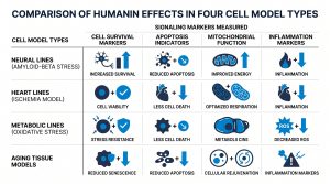
Most humanin cell studies focus on neural lines. The compound cuts markers of cell death when cells face amyloid-beta stress. This effect was seen in SH-SY5Y and PC12 cell lines.
According to a neural cell study (2017), humanin at nanomolar levels cuts caspase-3 activity by 40 to 60% versus control in amyloid-stressed cells. Caspase-3 is a key apoptosis marker. A drop in its activity means fewer cells enter the cell death program.
Humanin also affects tau protein levels in some models. Tau tangles are a marker of neural stress. Lab results show humanin cuts tau signal levels in amyloid-exposed cells. But the exact path for this effect is not yet clear.
How Is Humanin Used in Heart Cell Research?
Humanin has been studied in heart cell models for its effect on oxidative stress and energy output markers. Heart cells are high-demand energy users. They rely on healthy mitochondria for ATP. When mitochondria are damaged by oxidative stress, energy output drops and cell death signals rise.
Lab studies expose heart cell lines to ischemia-like conditions. Oxygen and glucose are removed, then restored. Humanin added before or during this step cuts cell death markers and keeps ATP output versus control groups. Results vary across cell lines and research setups.
Researchers using humanin in heart cell models should confirm lot purity before each study run.
How Does Humanin Compare to Other MDPs?
Humanin is one of several peptides encoded in mitochondrial DNA. MOTS-c is the most studied of the others. Both are MDPs but differ in size and receptor targets.
Humanin is 21 amino acids. MOTS-c is 16 amino acids. Humanin acts through CNTFR/WSX-1/gp130 on cell surfaces. MOTS-c acts through nuclear paths linked to gene output. In cell studies, humanin cuts apoptosis markers while MOTS-c alters glucose uptake and ATP output.
| Parameter | Humanin | MOTS-c |
| Length | 21 amino acids | 16 amino acids |
| Primary receptor | CNTFR, WSX-1, gp130 complex | Nuclear AMPK pathway |
| Main cell models | Neural lines, heart lines | Metabolic lines, muscle lines |
| Key signaling | JAK2/STAT3, PI3K/Akt | AMPK, PGC-1 alpha |
| Apoptosis effect | Reduces caspase-3 | Mixed, model-dependent |
| Purity standard | HPLC 99%+, mass spec | HPLC 99%+, mass spec |
What Are Supply Standards for Humanin Research?
Research-grade humanin must meet strict purity and records standards. HPLC purity must be at or above 99%. Mass spec must confirm the mass matches the 21-amino acid sequence. A lot-specific COA must link both results to a single run.
Key supply checks for researchers:
- HPLC purity at or above 99% on the COA.
- Mass spec check of correct molecular weight.
- Lot-specific COA, not a batch-level record.
- Outside lab testing alongside in-house testing.
- Freeze-dried form with residual moisture below 1%.
Every humanin lot from Next Level Pharm ships as a freeze-dried solid with a lot-specific COA. Every batch is third-party tested..
Frequently Asked Questions
What is humanin peptide?
Humanin is a 21-amino acid peptide encoded in mitochondrial DNA. It was first described in 2001 in neural cell research. Humanin binds to receptors on cell surfaces and starts JAK2/STAT3 and PI3K/Akt signal chains that control cell death and stress responses. In lab models, humanin is studied in neural, heart, and cell lines.
Where does humanin come from inside the cell?
Humanin is encoded in the 16S rRNA gene inside mitochondrial DNA. Most proteins made by mitochondria stay inside the cell part. Humanin is one of a small group that is sent out to act outside the cell. Cells release more humanin when mitochondria face oxidative or energy stress. This makes it a signal molecule that links mitochondrial state to cell behavior.
What receptors does humanin bind to?
Humanin binds to a three-part receptor complex on cell surfaces. The three parts are CNTFR, WSX-1, and gp130. All three must be present for full receptor activation. Binding triggers JAK2, which starts STAT3. STAT3 then moves into the nucleus and alters output of survival and apoptosis genes.
What signaling pathways does humanin activate?
Humanin starts two main pathways. JAK2/STAT3: JAK2 starts STAT3, which enters the nucleus and changes gene output. PI3K/Akt: PI3K makes a lipid signal that starts Akt to block pro-death signals. Both pathways cut apoptosis markers in treated cell lines. The two chains can work at the same time in most cell models studied.
What do neural cell studies show about humanin?
Neural cell studies show humanin cuts markers of cell death when cells face amyloid-beta protein. In SH-SY5Y and PC12 cell lines, humanin at nanomolar levels cuts caspase-3 activity by 40 to 60% versus control. Some studies also show a drop in tau signal levels in amyloid-stressed cells. The path for the tau effect is still under review in the research literature.
What do heart cell studies show about humanin?
Heart cell studies expose lines to ischemia-like conditions and then add humanin. Results show cut cell death markers and kept ATP output versus control groups. Humanin appears to cut oxidative stress signals through PI3K/Akt activation, which blocks pro-death signals. Results vary across cell lines and study setups, and need to be confirmed in each lab’s own model.
How does humanin differ from MOTS-c?
Humanin and MOTS-c are both mitochondria-derived peptides. But they differ in size and receptor targets. Humanin is 21 amino acids and binds to the CNTFR/WSX-1/gp130 surface complex. MOTS-c is 16 amino acids and acts through nuclear AMPK pathways. Humanin is mainly studied in neural and heart cell models. MOTS-c is mainly studied in muscle models for energy and glucose effects.
What purity is needed for humanin research?
Research-grade humanin needs HPLC purity at or above 99% and mass spec check of the correct 21-amino acid molecular weight. A lot-specific COA must link both results to one run. Studies using lower-purity humanin may show mixed results due to impurity signals in cell assays. Lot-specific COAs let researchers confirm exactly which vials were tested.
Where can researchers buy humanin peptide with COA data?
Next Level Pharm stocks research-grade humanin peptide as a freeze-dried solid. Each lot ships with a lot-specific COA that includes HPLC purity and mass spec results. Every batch is third-party tested.. This supports audit records for labs that track compound quality across multiple study runs.
Summary
Humanin is a 21-amino acid peptide encoded in mitochondrial DNA. It binds to a surface receptor complex and starts JAK2/STAT3 and PI3K/Akt signal chains. Both chains cut apoptosis markers in treated cell lines. Neural cell studies show humanin cuts caspase-3 activity in amyloid-stressed models. Heart cell studies show cut cell death markers and kept ATP output in ischemia models.
Humanin belongs to a group of mitochondria-derived peptides that includes MOTS-c. The two differ in size, receptor targets, and main study contexts. Both need HPLC purity above 99% and lot-specific COA records for valid research.
What Should You Do Next?
- Confirm the lot-specific COA shows HPLC purity above 99% and mass spec check before use in a study.
- Store freeze-dried vials at -20°C and bring to room temperature before opening.
- Review study setups for the cell line and humanin level used before your own assay.
Shop research-grade humanin peptide to get lot-specific HPLC and mass spec COA data with every order.
People Also Read
- MOTS-c Research: Mitochondrial Peptide and Metabolic Pathway Studies
- Recovery Peptides Compared: BPC-157, TB-500, KPV, and GHK-Cu Research
About the Author
Next Level Pharm Research Team
Alex M covers peer-reviewed findings in peptide science for Next Level Pharm, a US-based supplier of research-grade peptides verified to at least 99% purity via HPLC and mass spectrometry on every batch.
Disclaimer: The information provided on this page is for educational and research purposes only. Next Level Pharm’s products are intended for laboratory research use only. They are not intended for human consumption, diagnostic, therapeutic, or medicinal purposes. This content does not constitute medical advice. Always consult a licensed healthcare professional before making any health-related decisions