
Vasopressin Peptide Research: AVP Receptor Mechanism

Last updated: October 2026
A vasopressin AVP receptor peptide research study is an in vitro exam of how AVP, a nine-residue hormone, binds to its three GPCR subtypes. These subtypes are V1a, V1b, and V2, each linked to a distinct G protein and a separate signaling path. Mapping these binding events gives labs a framework for studying fluid balance, vascular tone, and stress signaling in cell models.
Next Level Pharm supplies lab-grade peptides for in vitro research. Each batch reaches a 99.4% average purity across the last 100 lots, confirmed by HPLC and mass spec. Lyophilized vials ship stable at room temperature, and a COA comes with every order.
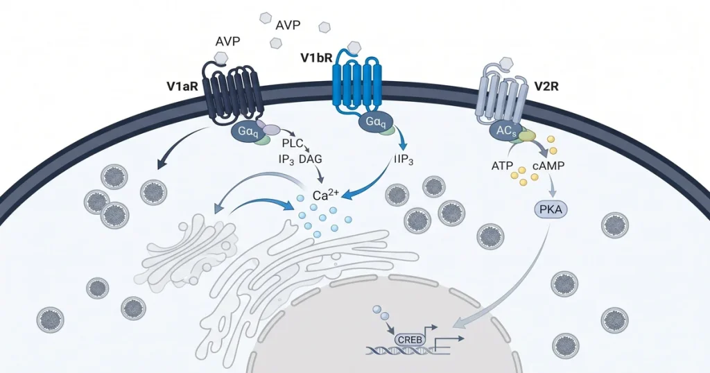
AVP signaling runs through three GPCR subtypes, each with a distinct G protein partner. The G protein choice sets whether the cell answers with calcium, cAMP, or another messenger.
Key Takeaways
- Peptide structure: AVP is a nine-residue neuropeptide with a disulfide ring and a short C-terminal tail.
- Three subtypes: V1a, V1b, and V2 each link to a different G protein and fire a distinct cell path.
- Calcium path: V1a and V1b couple to Gq/11 to fire PLC, which raises calcium in the cell.
- Water path: V2 couples to Gs to raise cAMP, then PKA moves AQP2 to the collecting duct.
- Assay methods: Labs use binding assays and calcium or cAMP functional assays in HEK293 or CHO cells.
- Selective blockers: Vaptans are nonpeptide AVP blockers that target one subtype at a time in cell models.
The sections below cover each mechanism with lab data. Each section builds on the receptor type and G protein pair.
What Is Arginine Vasopressin (AVP)?
AVP is a nine-residue neuropeptide made in the hypothalamus and stored in the brain’s posterior gland.
Rising plasma osmolality or low blood volume triggers AVP release into the blood.
Once in the blood, AVP binds to GPCR subtypes on kidney, vascular, and gland cells.
AVP shares close structural features with oxytocin. The two peptides differ at only two amino acid positions.
According to a PubMed review on AVP receptor physiology (2008), both peptides share ancestral origins and can weakly bind each other’s GPCR sites in vitro.
How Do Vasopressin Receptors Signal?
AVP receptors are class A GPCRs that span the cell membrane via seven helices.
When AVP binds, the receptor shifts shape and activates a linked G protein.
The G protein type sets which second messenger fires next.
- V1a and V1b: Gq/11-linked, raise calcium via PLC in smooth muscle, platelet, and gland cells.
- V2: Gs-linked, raises cAMP via AC in kidney collecting duct cells.
- Signal output: Each subtype produces a distinct response based on G protein class.
According to a PMC receptor binding study (2017), CHO cells expressing human V1 or V2 GPCRs allow direct comparison of calcium and cAMP outputs. This setup lets labs see how small ligand changes shift receptor choice.
What Are V1a and V1b Receptor Functions?
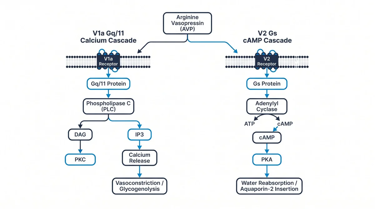
V1a and V1b both link to Gq/11 proteins in the cell membrane.
Gq/11 activation fires PLC, which makes IP3 and DAG.
IP3 then releases calcium from the ER into the cell.
The V1a receptor sits on smooth muscle cells, platelets, and liver cells. Calcium release drives muscle tightening and platelet clumping in these tissues. The V1b receptor sits on pituitary corticotroph cells, where calcium drives ACTH release.
Labs sourcing lab-grade peptides for V1a and V1b studies can browse the recovery peptide catalog at Next Level Pharm.
How Does the V2 Receptor Regulate Water?
The V2 receptor links to Gs. Gs activates AC, which converts ATP to cAMP. Rising cAMP then fires PKA.
PKA phosphorylates AQP2 water channels stored in cell vesicles inside the duct.
This step moves AQP2 to the apical membrane, raising water flow across the cell.
According to a PMC study on AQP2 translocation (2025), AQP2 density at the apical membrane is set by a reversible AVP-driven recycling cycle.
How Is AVP Receptor Activity Measured?
Labs measure binding affinity with a labeled tracer ligand and cell membrane fractions.
The tracer competes with the test compound for GPCR sites.
Binding data fit a sigmoidal curve, yielding a Ki value.
- HEK293 cells: Low GPCR background; preferred for calcium or cAMP functional screens.
- CHO cells: High membrane yield; preferred for tracer binding assays.
- Ki value: Ranks test compound potency at one receptor subtype per assay run.
For V1a and V1b, labs measure calcium flux using a dye-based plate assay. For V2, labs measure cAMP buildup using HTRF or enzyme-linked kits. Both methods run in 96-well or 384-well plate formats.
What Are Vasopressin Receptor Antagonists?
Vaptans are nonpeptide AVP blockers that occupy the GPCR binding pocket without firing a signal.
They let labs remove AVP signaling from a model while all other systems stay active.
This makes vaptans key tools for isolating each subtype’s role.
Conivaptan blocks both V1a and V2. Tolvaptan blocks only V2. Research-grade Arginine Vasopressin and vaptan analogs are COA-confirmed compounds for in vitro lab use.
How Do AVP Receptor Subtypes Compare?
| Feature | V1a | V1b | V2 |
|---|---|---|---|
| G protein | Gq/11 | Gq/11 | Gs |
| Second messenger | Ca2+ via IP3 | Ca2+ via IP3 | cAMP |
| Main tissue | Smooth muscle, liver | Pituitary gland | Kidney duct |
| Key output | Muscle tightening | ACTH release | AQP2 translocation |
| Selective blocker | SR49059 | SSR149415 | Tolvaptan |
| Common cell line | CHO-V1a | HEK293-V1b | HEK293-V2 |
Frequently Asked Questions
What are the V1a and V2 vasopressin receptor subtypes?
The V1a receptor is a Gq/11-linked GPCR found on smooth muscle cells, platelets, and liver cells.
It fires the PLC path, which makes IP3 and raises calcium in the cell.
This calcium response drives muscle tightening and platelet clumping in tissue models.
The V2 receptor is a Gs-linked GPCR found in kidney collecting duct cells.
It raises cAMP, fires PKA, and moves AQP2 channels to the cell surface.
How does AVP bind V1R versus V2R?
AVP presents its nine-residue chain to both V1R and V2R.
Each receptor reads the same ligand but couples to a different G protein.
This G protein choice sets the downstream cell path.
V1R links to Gq/11 and starts a calcium response in smooth muscle and liver cells. V2R links to Gs and starts a cAMP response in kidney collecting duct cells. The two paths differ in output type, cell location, and downstream effect.
What signaling cascades do vasopressin receptors activate?
V1a and V1b both link to Gq/11 proteins in the cell membrane.
This step fires PLC, which makes IP3 and DAG as second messengers.
IP3 then triggers calcium release from the ER into the cell.
V2 links to Gs, which fires AC to produce cAMP. Rising cAMP activates PKA, which phosphorylates AQP2 channels. Each path has its own timing and output level.
How is vasopressin receptor binding affinity measured?
Binding affinity for each AVP receptor subtype is measured with a tracer competition assay.
A labeled AVP analog is mixed with receptor-expressing cell membranes at a set amount.
The test compound is then added at rising levels to displace the tracer.
Binding data fit a sigmoidal curve to yield a Ki value. This Ki reflects the compound’s potency at that subtype.
What cell lines express vasopressin receptors for research?
HEK293 and CHO cell lines are the most common hosts for AVP receptor research.
Both lines accept stable insertion of V1a, V1b, or V2 receptor constructs.
Labs choose them for their reliable growth and consistent membrane yields.
HEK293 cells have low background GPCR activity, giving clean calcium or cAMP readouts. CHO cells produce high membrane protein yields, making them ideal for tracer binding screens.
What is the difference between vasopressin and desmopressin?
AVP is the native nine-residue hormone that activates V1a, V1b, and V2.
Desmopressin is a synthetic AVP analog with two key structural changes.
One change removes the N-terminal amino group; the other swaps L-Arg at position 8 with D-Arg.
These changes make desmopressin selective for V2, cutting V1a-driven effects. This makes it a standard tool for isolating V2 renal water signaling in cell models.
How is synthetic vasopressin produced for lab studies?
Synthetic AVP is made by solid-phase peptide synthesis (SPPS).
SPPS builds the nine-residue chain one amino acid at a time on a solid resin.
The method uses Fmoc or Boc chemistry to add each protected residue in order.
After full assembly, oxidative folding forms a disulfide bridge between Cys-1 and Cys-6. HPLC and mass spec then confirm purity before the batch is lyophilized.
Are vasopressin and oxytocin structurally related?
AVP and oxytocin are both nonapeptides with a six-residue disulfide ring and a three-residue tail.
They share seven of nine amino acid positions.
The two differ at positions 3 and 8 only.
In AVP, position 3 holds Phe and position 8 holds Arg. In oxytocin, Ile sits at 3 and Leu sits at 8. This two-residue gap lets each peptide weakly bind the other’s GPCR sites at high levels in vitro.
What are vaptans and how are they used in lab work?
Vaptans are nonpeptide AVP GPCR blockers that halt hormone binding without triggering a signal.
Labs apply vaptans to cell models to silence one receptor subtype at a time.
This isolates each subtype’s share of the total AVP response.
Conivaptan blocks both V1a and V2, while tolvaptan blocks V2 only. Comparing treated and control cells shows which outputs come from V1a versus V2.
What does V1b receptor activation control in research models?
V1b activation drives ACTH release from pituitary corticotroph cells.
ACTH then signals the adrenal gland to release cortisol.
This places V1b at the center of the HPA axis in stress signaling models.
Cell lines that express V1b let labs study pituitary hormone output in isolation from V1a and V2 events. Selective V1b blockers are standard tools for HPA axis research in these models.
Summary
AVP is a nine-residue neurohormone that signals through three GPCR subtypes. V1a and V1b couple to Gq/11 to raise Ca2+ via PLC. V2 couples to Gs to raise cAMP and move AQP2 to the collecting duct.
Standard methods include tracer binding assays for affinity and cell-based functional assays in HEK293 or CHO lines.
Vaptans let labs block individual subtypes and isolate their specific signaling roles.
Research-grade AVP, desmopressin, and vaptan analogs are COA-confirmed compounds. Next Level Pharm stocks them as pure, lab-tested peptides for in vitro experiments.
What Should You Do Next?
Labs starting AVP receptor work should confirm receptor subtype expression in the chosen cell line. Western blot or flow analysis can verify expression levels before assays begin.
Researchers sourcing peptides for AVP receptor work can browse the full research peptide catalog at Next Level Pharm. Every peptide ships with a COA, HPLC trace, and mass spec confirmation at the stated lot number.
People Also Read
- Surface Plasmon Resonance Peptide Binding Research
- MAPK ERK Pathway Peptide Research: Kinase Cascade
About the Author
Next Level Pharm Research Team
The Next Level Pharm research team is composed of biochemists and laboratory scientists dedicated to providing researchers with the highest-purity, COA-tested peptides available. Every batch is HPLC and mass spec confirmed before dispatch.
Disclaimer: The information provided on this page is for educational and research purposes only. Next Level Pharm’s products are intended for laboratory research use only. They are not intended for human consumption, diagnostic, therapeutic, or medicinal purposes. This content does not constitute medical advice. Always consult a licensed healthcare professional before making any health-related decisions.