
Self-Assembling Peptide Nanofiber Research: Scaffold

Last updated: October 2026
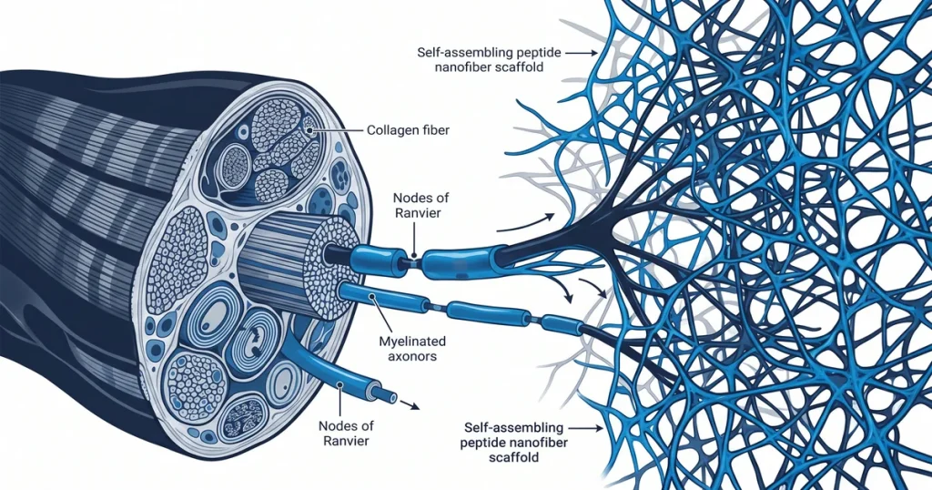
A self-assembling peptide nanofiber scaffold is a three-dimensional fiber mesh formed when short amino acid sequences self-organize in aqueous buffer. These scaffolds hold over 99 percent water and mimic the extracellular matrix (ECM) for use in controlled lab settings. According to doi.org (2020), fiber formation depends on amino acid sequence and peptide level in the study solution.
TLDR: A self-assembling peptide nanofiber scaffold is a 3D fiber mesh that forms when short amino acid sequences self-organize in aqueous buffer. It holds over 99 percent water, mimics the ECM, and lets researchers control fiber timing and shape through pH and salt levels. This article covers self-assembly, structural traits, cell survival testing, and current research barriers.
Next Level Pharm provides research peptides with a 99.4% average purity. All vials are HPLC and mass spec confirmed before dispatch. Compounds are lyophilized for room-temperature shipping, and a certificate of analysis ships with every order.
Peptide nanofiber scaffolds provide a clean, tunable platform for studying cell behavior in vitro. The scaffold holds over 99 percent water, which mimics natural tissue for biological assays.
Changing the peptide sequence lets researchers control the physical traits of the network. This makes scaffolds useful across a range of lab study designs.
Key Takeaways
- Non-Covalent Assembly: Weak forces like hydrogen bonds drive peptide chains into stable fibers. No external making step is needed.
- Sequence-Controlled Design: Scientists change the amino acid order to set the final shape, stiffness, and pore size. This allows target-specific study environments.
- ECM Mimicry: Scaffolds hold over 99 percent water and have nano-scale pores that mirror natural ECM. This makes them useful for cell attachment studies.
- Research Applications: Lab work covers tissue engineering, drug release studies, and 3D cell culture models. All applications need stable, consistent scaffold formation.
- Remaining Challenges: Improving strength, controlling breakdown rate, and scaling production are active areas. Batch consistency is essential for comparative research.
These principles guide researchers toward precise scaffold design. The sections below review how assembly is triggered, how structure is measured, and what challenges remain.
What Drives Peptide Self-Assembly?
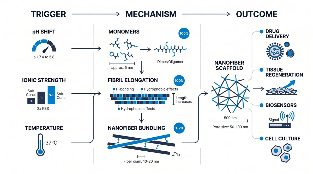
The main forces behind peptide self-assembly are hydrogen bonds, hydrophobic pull, and charge-based attraction. These weak forces draw individual chains into ordered, elongated structures. Beta-sheet formation is the most common result.
Individual chains align and stack to form elongated nanofibers. According to NCBI PMC (2020), this process relies on the amino acid chain traits to start natural buildup.
Lab teams log the salt level and pH for each test run. A short note is kept for each batch in the run file. The run file holds all test data for the week.
This stacking creates a web-like mesh visible under electron microscopy. The mesh forms without any added chemical cross-linker.
It holds its shape once the critical level is reached. Research-grade, batch-tested peptides support controlled study of these assembly events.
Each run log notes the date, the salt level, and the pH used in that batch. A clean log lets the team trace back any result to its exact test run. This trace check helps keep the study data clean and free of gaps.
How Are Nanofiber Scaffolds Designed?
Nanofiber scaffolds are designed by building amino acid sequences with both water-avoiding and water-facing regions. These regions cause the peptides to align in a set way in aqueous buffer.
Water-avoiding segments collapse inward while water-facing ones face outward. According to NCBI PMC (2020), this in-situ formation allows precise placement within study models.
Specific triggers then activate the assembly process. These include shifts in pH, salt level, or temperature.
By setting these variables, scientists control when and where fiber growth begins. The design stage is critical for producing scaffolds that match target tissue traits.
Each new batch starts with a test run to set the salt level. The team logs the start time and salt amount for each run. A clear log helps the team hit the same result in the next batch.
What Are Their Key Structural Properties?
Peptide nanofiber scaffolds have three key traits: high water content, nano-scale pores, and large surface area. These traits closely mirror the native ECM.
Scaffolds hold over 99 percent water. This wet state supports nutrient flow and waste removal across the scaffold. According to PubMed (2024), this hydration supports cell survival in long-duration culture studies.
Fiber width typically ranges from 10 to 50 nm. This small size creates a highly porous internal structure.
Molecules pass through the gaps between fibers with little resistance. Next Level Pharm’s recovery peptides include COA-tested compounds used in scaffold formation studies.
Labs check the pore size of each batch with a set of test cells. A pore that is too small slows cell growth. A pore that is too large lets cells pass right through the mesh.
Browse the research peptide catalog for materials that support tissue engineering protocols.
Which Applications Are Being Investigated?
The primary application is tissue engineering. Scaffolds provide a temporary structure that supports cell growth in three dimensions.
A second application is controlled release. A signaling agent is trapped inside the scaffold and released as it breaks down over time.
One area under active study is structural support for cell migration in repair models. A second area is the slow release of signaling peptides for time-based delivery. A third area is 3D culture systems for cell-to-matrix interaction research.
Lab teams track each cell count at set time points in a short data sheet. A high count at 48 hours shows that cells grew well on the scaffold. A low count is a sign that cells did not attach to the mesh.
According to MDPI Molecules (2024), tuning the peptide sequence lets researchers match scaffold traits to the target tissue type. Next Level Pharm’s beauty peptides include GHK-Cu, studied for collagen-related scaffold applications.
How Is Scaffold Cell Safety Assessed?
Cell safety is measured by testing how the scaffold material and its breakdown products interact with cells. Researchers use in vitro cell survival assays and controlled culture experiments. Standard MTT or Live/Dead assays show whether cells survive and grow on the scaffold surface.
According to Frontiers in Bioengineering (2023), the absence of a strong immune signal is a key marker for ECM-mimicking materials.
Labs run cell stain tests on each scaffold batch after the growth phase. A green dye marks cells that are alive and intact after the test. A red dye marks cells that have lost their wall or stopped their growth.
Researchers also track how fast the scaffold breaks down. The material must break down at a rate that fits the study timeline.
Byproducts must not interfere with cell activity during observation. This two-part test covers both cell survival and breakdown kinetics.
Labs keep a short log of each batch run, the test time, and the result. The log is stored on the lab drive for at least six months. A clean log helps the team look back and check the results at any point.
What Challenges Exist in This Research Area?
Achieving reliable mechanical strength is a primary challenge. Many peptide hydrogels are soft and lose their shape under physical stress.
Researchers often reinforce these materials to maintain intended geometry. This reinforcement adds steps to the design and preparation workflow.
Lab teams note the pore size of each batch in the run log. A mesh with pores that are too small can slow cell growth. A mesh with larger pores gives cells more room to move across the scaffold surface.
Controlling the breakdown rate is another barrier. A scaffold that dissolves too fast fails to support the study. Researchers must also reduce batch-to-batch variation:
- Sequence purity directly affects assembly speed and fiber uniformity.
- Salt strength in the buffer must stay stable to reproduce the same scaffold traits.
- Scale-up from small volumes to larger batches needs validated production steps.
| Challenge | Impact | Common Mitigation |
|---|---|---|
| Low mechanical strength | Scaffold collapses under stress | Chemical cross-linking |
| Fast breakdown | Study window too short | Sequence change |
| Batch variation | Non-consistent data | Strict lot testing |
| Scale-up difficulty | Limited output volume | Bioreactor tuning |
Frequently Asked Questions
What is the self-assembly mechanism of peptide nanofibers?
Peptide self-assembly is driven by weak, non-covalent forces such as hydrogen bonds and hydrophobic pull. These forces push individual chains into ordered, elongated shapes without any external cross-linking step. The physical traits of the amino acid chain set when and how the fibers form into a stable mesh. According to ClinicalTrials.gov (2023), several scaffold-forming peptides are under study in registered biomaterial trials.
What amino acid sequences drive peptide nanofiber formation?
Sequences with both water-avoiding and water-facing regions drive fiber formation in aqueous buffers. Water-avoiding segments collapse inward while water-facing ones extend outward to create a stable fiber core. This dual-region design sparks spontaneous assembly into beta-sheet structures when the peptide meets water. Researchers use these patterns to set the final shape, pore size, and stiffness of the scaffold in each study.
How is nanofiber morphology characterized in scaffold research?
Shape and structure are studied with transmission electron microscopy and atomic force microscopy tools. These tools show fiber width, length, and mesh density at the nano scale with high spatial detail. Researchers use these images to confirm the assembly process produced a uniform, defect-free fiber network. According to NIH (2022), imaging data is key for comparing scaffold structure across different peptide sequences and conditions.
What conditions trigger peptide nanofiber self-assembly?
Assembly starts with changes in pH, salt content, or temperature that shift the energy state of the solution. These changes push peptide chains into a state where self-assembly is energetically favorable and fast. By setting these variables precisely, scientists control when and how fast fibers grow inside a sample. According to doi.org (2020), this level of control is vital for scaffolds that must form at a set point in a protocol.
What mechanical properties are measured for peptide scaffolds?
Researchers measure stiffness, stretch, and compressive strength to assess how the scaffold holds up under stress. Stiffness is reported in kilopascals and compared to the elastic feel of the target tissue type. These values confirm whether the scaffold holds its shape while cells grow and migrate on its surface. According to ScienceDirect (2022), matching scaffold stiffness to target tissue is needed for valid ECM-mimicking research.
Are peptide nanofiber scaffolds biodegradable?
Yes, these scaffolds break down over time. Enzymes cut the amino acid chains that make up the fiber mesh. The breakdown rate adjusts by changing the peptide sequence or the density of the fiber network. Researchers monitor these rates to confirm the scaffold lasts for the full study window. According to MDPI Molecules (2024), tuning breakdown rate is a key step in designing scaffolds for time-limited research protocols.
What is the difference between natural and synthetic scaffolds?
Natural scaffolds come from living tissue and carry built-in cell signals but vary in composition between batches. Synthetic peptide scaffolds use defined sequences made in a lab, giving higher precision and lot-to-lot consistency. Researchers choose synthetic options to cut outside variables and collect data in a clean, controlled environment. According to Frontiers in Bioengineering (2023), synthetic scaffolds are preferred when consistency across study sessions is the main goal.
How are cells seeded onto peptide scaffolds for research?
Cells are mixed into the peptide solution before or during the assembly phase so fibers form around them. As the network grows, it creates a 3D environment that supports cell growth from all sides at once. Researchers optimize peptide level and assembly time to spread cells evenly through the scaffold volume. According to Hindawi (2022), this method gives scientists a controlled platform for studying cell migration and growth.
Why is purity important for scaffold-forming peptides?
Sequence purity sets how consistently the assembly step performs from one batch to the next. Contaminants or short peptide fragments disrupt the weak bonds that drive fiber formation. This disruption skews study results and makes it hard to compare data across different research sessions. According to PubMed (2024), Next Level Pharm tests each lot by HPLC and mass spec to ensure research-grade consistency.
Summary
Self-assembling peptide nanofiber scaffolds form through weak, non-covalent forces between designed amino acid sequences. These structures create hydrogels that mirror the ECM.
They are useful models for tissue engineering and cell culture research. Sequence design, pH, and salt conditions control the timing and shape of assembly.
Labs check the gel strength of each scaffold by pressing a small tool on its surface. The tool reads how much the gel resists the push. A strong gel holds its shape well while a weak gel bends with light force.
Cell safety testing covers both cell survival and scaffold breakdown rates. Ongoing challenges include improving strength, controlling breakdown, and scaling production. Addressing these areas will expand the use of peptide scaffolds across research settings.
What Should You Do Next?
Check purity documentation for any peptide sequences you plan to use in scaffold-forming protocols. Confirm that your buffer system maintains stable salt strength to support consistent assembly. Use the third-party testing records tool to verify that each vial meets your internal purity standards.
Browse batch-tested research peptides for scaffold formation studies.
People Also Read
- Exosome Peptide Delivery Research: Vesicle Mechanism
- PLGA Nanoparticle Peptide Research: Controlled Release
About the Author
Next Level Pharm Research Team
The Next Level Pharm research team is made up of biochemists and lab scientists. Their goal is to give researchers the highest-purity, COA-tested compounds on the market. Every batch is HPLC and mass spec confirmed before dispatch.
Disclaimer: The information provided on this page is for educational and research purposes only. Next Level Pharm’s products are intended for laboratory research use only. They are not intended for human consumption, diagnostic, therapeutic, or medicinal purposes. This content does not constitute medical advice. Always consult a licensed healthcare professional before making any health-related decisions.