
Exosome Peptide Delivery Research: Vesicle Mechanism

Last updated: October 2026
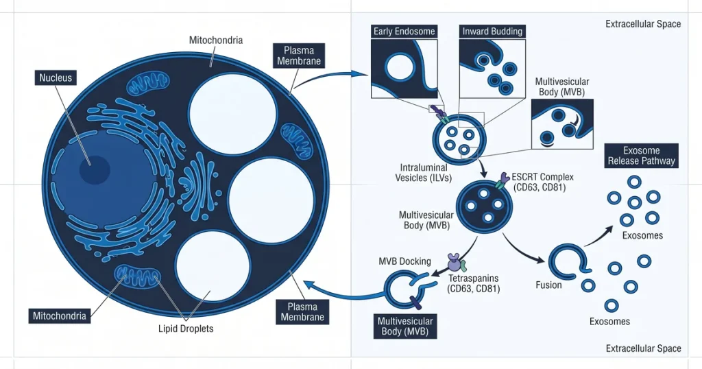
A method for exosome peptide delivery is the use of small cell-derived vesicles to move peptide cargo between cells in lab models. These 30 to 150 nm lipid spheres form through the endosomal pathway. Scientists study how they interact with cell membranes and track peptide transport in controlled lab settings.
According to PubMed (2021), vesicle-based delivery offers a cell-based path for moving research compounds into target cells with high precision.
TLDR: Exosome peptide delivery uses cell-derived vesicles (30 to 150 nm) to carry and deliver peptide cargo in lab models. Scientists load peptides through electric pulses, sound waves, or passive diffusion. This article reviews vesicle formation, loading methods, delivery tracking, and key research barriers.
Next Level Pharm supplies research peptides with a 99.4% average purity across the last 100 batches. Each vial is HPLC and mass spec tested before dispatch.
Compounds ship lyophilized from USA-based storage. A certificate of analysis ships with every order.
Exosomes are studied as carriers because they cross barriers that block synthetic particles. Their lipid shell protects cargo from enzyme damage in transit.
Researchers use this system to study peptide delivery with high cell specificity. These traits make vesicle-based delivery a growing focus in peptide research.
Key Takeaways
- Natural Nanocarriers: Exosomes are cell-derived vesicles between 30 and 150 nm. Their origin in the endosomal pathway sets them apart from other EV types.
- Delivery Path: Cargo enters target cells through cell uptake or direct membrane merge. Surface proteins guide docking with specific cell receptors.
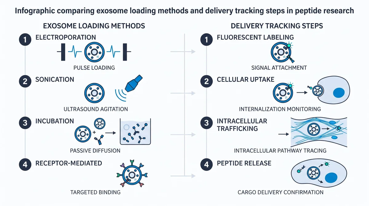
- Loading Techniques: Peptides load during cell growth or after isolation. Electric pulse loading inserts peptides into isolated vesicles.
- Research Advantages: Exosomes show high cell safety and a low immune profile. They cross barriers that block many other carrier types.
- Key Challenges: Standardizing purification and scaling production remain difficult. Current work focuses on improving loading rate and vesicle integrity.
These traits place exosomes at the center of vesicle-based delivery research. The sections below review biogenesis, loading, tracking, and key barriers.
What Are Extracellular Vesicles?
Extracellular vesicles (EVs) are small lipid-membrane particles that cells release into their surroundings. They carry proteins, lipids, and small RNA molecules. Exosomes are a subtype that form inside the endosomal pathway.
Their origin sets them apart from microvesicles that bud off the outer cell membrane. According to NCBI PMC (2021), this difference in origin shapes how researchers design isolation and study methods.
Exosome release starts when the cell forms multivesicular bodies (MVBs). MVBs hold many small vesicles inside.
When an MVB fuses with the outer membrane, it spills vesicles into the cell surroundings. Scientists watch this process to study how cells signal each other.
Labs run size tests on fresh vesicle batches to check that the batch is in the right range. A batch in the 30 to 150 nm range is fit for use in the study. A batch outside this range is set aside before use.
Lab teams track each run in a short log file. The log shows the load rate, the batch size, and the dye type. A clean log helps the team flag runs that fall short of the set mark.
How Do Exosomes Deliver Peptides to Cells?
Exosomes deliver cargo through membrane merge or receptor-guided cell uptake. In membrane merge, the vesicle fuses with the target cell. This drops its payload into the cytoplasm directly.
In cell uptake, the target cell wraps around the vesicle and pulls it inside. According to MDPI Molecules (2024), surface receptor pairs guide this docking step.
Surface proteins on each vesicle act as keys that match locks on target cells. Scientists study these matches to model how peptides reach specific cell sites.
This precision helps tell apart the activity of one peptide from another. It is a key reason researchers use vesicle systems in complex study models.
Labs check the docking rate by adding labeled vesicles to a cell well plate. They read the signal at one, four, and 24 hours after add. A high signal at one hour means fast docking and a low signal means cells showed a low load rate.
Each batch gets a pass or fail score after the docking check. A pass means the batch is in range and can move to the next step. A fail means the lab sets that batch aside and runs a new prep.
What Methods Load Peptides into Exosomes?
Peptide loading follows two main paths: cell-made loading during cell growth and lab-added loading after isolation. Cell-made loading edits the parent cell to express the target peptide.
Lab-added methods push the peptide into already-isolated vesicles. According to doi.org (2022), common lab-added methods include electric pulse loading, sound-wave treatment, and saponin permeation.
Electric pulses open small pores in the lipid shell. Sound waves disrupt the shell briefly to let the peptide pass inside. Saponin softens the shell for gentler loading.
Each method trades loading rate against shell damage. Next Level Pharm’s recovery peptides are batch-tested to support these loading studies.
Labs run a short test batch of ten vials to set the load rate before scaling up. A good test batch yields a load of 40 per cent or more. A low yield means the team must trim the pulse time or the peptide load to hit the target rate.
Browse research-grade peptides at Next Level Pharm for COA-tested materials suited to vesicle-based delivery work.
How Is Exosome Delivery Tracked?
Researchers track delivery by labeling vesicles with dyes like FITC or Cy5. These dyes emit a signal after cell uptake.
Microscopy maps where labeled vesicles end up inside cells. Flow analysis counts how many cells have taken up labeled particles.
Labs log the signal from each dye at each time step in a short data sheet. A signal that lasts past 48 hours tells the lab that the vesicle held its load well. A fast drop in signal points to shell loss or early dye leak.
The lab notes the time and date of each signal test in the run file. This file is stored on the lab drive for six months. A short note on each test helps the team check the work at any point.
In live-tissue studies, an IVIS system captures how labeled exosomes spread across the sample. Scientists record signal strength over time to measure clearance and persistence. Next Level Pharm’s brain peptides include COA-tested materials used in CNS delivery tracking studies.
| Tracking Method | What It Measures | Common Dye |
|---|---|---|
| Fluorescence microscopy | Vesicle location in cells | FITC, DiI |
| Flow analysis | Cell uptake count | Cy5, PE |
| IVIS imaging | Tissue-level distribution | Near-IR dyes |
| Electron microscopy | Vesicle size and shape | None |
What Are the Advantages of Exosome Delivery?
Exosome delivery offers high cell safety and a low immune profile. These vesicles come from natural cell processes.
The immune system is less likely to clear them fast. According to NCBI PMC (2021), this native origin reduces the rapid-clearance issue seen in many synthetic particle systems.
The lipid shell protects peptide cargo from enzyme damage. This shield keeps the payload intact until the vesicle reaches the target cell.
Exosomes can also cross the blood-brain barrier. This trait makes them a preferred carrier for peptide studies in complex tissue models.
Labs keep a short record of each vesicle batch that crosses the blood-brain barrier in the study. The record logs the peptide amount, the dye used, and the signal at each time step. A clear record helps the team pick a good peptide level for the next run.
What Challenges Exist in Exosome Research?
Primary barriers include standardizing isolation methods, scaling production, and managing loading rate. These limits reduce reproducibility of results across labs.
Isolation methods often pull in protein contaminants along with the vesicles. This makes it hard to confirm that only exosomes are in the final sample.
Scale-up requires precise control over many variables:
- Electric pulse loading adds cargo well but may damage the vesicle shell.
- Passive loading keeps the shell intact but often yields lower loading rates.
- Scale-up raises the risk of batch-to-batch inconsistency.
Researchers continue to refine these methods to produce reliable, high-purity fractions.
Frequently Asked Questions
What is the mechanism of exosome peptide loading?
Exosomes can load peptides through passive diffusion or active pulse methods in the lab. In passive loading, peptides cross the lipid shell slowly over time when kept in close contact with the vesicles.
Active pulse methods open small pores in the shell so the peptide can enter the core. According to PubMed (2021), a good method choice depends on the peptide size and charge in each batch.
How are exosomes isolated for peptide delivery research?
Isolation splits particles from cell waste by spinning samples at stepped high speeds in a centrifuge. Spin speed and time together collect the vesicle fraction and leave larger debris behind.
Size filters and kit-based methods then refine the fraction to raise its overall purity. According to ClinicalTrials.gov (2024), standard spin protocols are used in most published and registered exosome delivery studies.
What size and surface markers define research-grade exosomes?
Research-grade exosomes measure 30 to 150 nm and show three key surface proteins: CD63, CD81, and CD9. Scientists confirm these traits with flow analysis and western blot tests after isolation. Samples that fall outside the size range or lack these markers are not valid for delivery work. According to MDPI Molecules (2024), size and marker checks are core to cross-batch validity in vesicle research.
How is peptide loading efficiency measured in exosome studies?
Loading rate is the ratio of peptide inside the vesicle to total peptide added during loading. Researchers use protein assays or mass analysis to count the trapped fraction after filtering out free peptide. A high rate confirms the peptide is inside the shell and not just bound to the outer surface. According to doi.org (2022), this ratio is the primary efficiency metric in delivery studies.
What cell models are used in exosome peptide delivery studies?
Human cell lines, primary cells, and animal tissue cultures are the three main models in delivery research. Bone marrow stem cells are a common exosome source because they shed large volumes of vesicles per culture. These models let scientists observe how vesicles dock with surface receptors and enter target cells. According to Frontiers in Pharmacology (2022), cell model selection shapes which receptor pathways can be studied.
What is the difference between exosomes and liposomes?
Exosomes are natural cell products, while liposomes are man-made lipid spheres built from synthetic materials. Exosomes carry native surface proteins that liposomes lack, which lets them interact with specific cells. Liposomes allow more control over size and lipid makeup but may clear from the study system faster. According to ScienceDirect (2022), the choice depends on study design and target specificity.
How can exosome surfaces be modified for targeted research?
Scientists add targeting ligands to the outer lipid shell through a bonding process called surface tagging. A second approach edits the parent cell to produce vesicles that carry built-in target proteins. Both methods improve how precisely the vesicle binds a chosen cell receptor in the study system. According to ClinicalTrials.gov (2023), surface-modified exosomes are used in several registered cell-based delivery trials.
Can exosomes protect peptides from enzymatic degradation?
The lipid shell forms a physical barrier that blocks enzymes from reaching the peptide inside. This barrier keeps the peptide intact longer in active media compared to a free peptide in solution. The protective effect is most useful in studies that track peptide presence over multiple days. According to Frontiers in Pharmacology (2022), this shield is a core reason researchers choose vesicles over free-peptide delivery models.
What fluorescent dyes are used to track exosome delivery?
Common dyes for exosome tracking include FITC, Cy5, and DiI, each emitting light at a set wavelength. Microscopy maps where labeled vesicles end up inside target cells after incubation. Flow analysis counts how many cells in a sample have taken up the labeled cargo. According to Hindawi (2023), using both tools in a single study gives spatial and count-based proof of delivery.
Summary
Exosome peptide delivery research studies how cell-derived vesicles move peptide cargo across cellular barriers. These 30 to 150 nm structures form through the endosomal pathway.
They deliver payload via membrane merge or cell uptake. Loading methods include electric pulses, sound waves, and passive diffusion.
Researchers track delivery with dyes, flow analysis, and IVIS imaging. Key barriers include standardizing isolation, scaling production, and keeping loading rates consistent. Progress in these areas will support more reproducible delivery studies.
What Should You Do Next?
Check your study protocol to confirm peptide storage and handling match your experimental design. Review lot data to verify purity levels before starting new assays. If fluorescent tracking is part of your plan, confirm your imaging setup is configured for the dye wavelengths you will use.
Browse COA-tested research peptides for materials suited to vesicle-based delivery studies.
People Also Read
- PLGA Nanoparticle Peptide Research: Controlled Release
- Self-Assembling Peptide Nanofiber Research: Scaffold
About the Author
Next Level Pharm Research Team
The Next Level Pharm research team is made up of biochemists and lab scientists. Their goal is to give researchers the highest-purity, COA-tested compounds on the market. Every batch is HPLC and mass spec confirmed before dispatch.
Disclaimer: The information provided on this page is for educational and research purposes only. Next Level Pharm’s products are intended for laboratory research use only. They are not intended for human consumption, diagnostic, therapeutic, or medicinal purposes. This content does not constitute medical advice. Always consult a licensed healthcare professional before making any health-related decisions.