
Peptide Research in Gut Mucosal Biology

Last updated: July 2026
A peptide gut mucosal biology study is a controlled lab model that tests how small protein chains interact with the intestinal lining. The gut mucosa is the inner lining of the intestine. It is made of gut cells, a mucus layer, and an underlying layer of connective tissue. Peptides like BPC-157 and KPV are studied for how they change cytokine output and repair signals in gut cell cultures. According to PubMed (2019), BPC-157 adjusts cytokine balance in mucosal tissue models.
Next Level Pharm supplies research-grade gut mucosal peptides for lab use. Every vial is HPLC-tested and ships with a COA (certificate of analysis). Average purity across recent batches is 99.4%.
Gut mucosal biology research uses peptides to probe how the intestinal lining responds to chemical signals. The mucosa acts as a barrier between the gut contents and the bloodstream. When barrier function drops, immune cells become more active. Peptides that support mucosal integrity or lower cytokine output are key tools in this research area.
Key Takeaways
- Mucosal Barrier Is the Central Target: The gut mucosal barrier (the single-cell-thick lining of the intestine) controls what enters the bloodstream. Peptides that support barrier function are a primary focus in gut mucosal biology.
- BPC-157 in Mucosal Repair Models: BPC-157 is a 15-amino-acid peptide. It is studied for its effects on gut epithelial repair, cytokine balance, and nitric oxide output in mucosal lab models.
- KPV as an Epithelial Anti-Inflammatory Tool: KPV (Lys-Pro-Val) blocks NF-kB signal in gut cells. This lowers pro-inflammatory cytokines in gut cell culture models.
- GHK-Cu and Connective Tissue Support: GHK-Cu (copper peptide) is studied for collagen production and extracellular matrix repair in gut tissue assays.
- TB-500 and Mucosal Cell Migration: TB-500 (thymosin beta-4 analog) promotes gut cell movement to close tissue gaps in mucosal wound healing models.
- COA-Verified Gut Mucosal Peptides: All gut mucosal research peptides listed here are lyophilized and batch-tested. COA records link to each lot number for full lab traceability.
These six points define the research landscape covered in the sections below.
What Is Gut Mucosal Biology in Peptide Research?
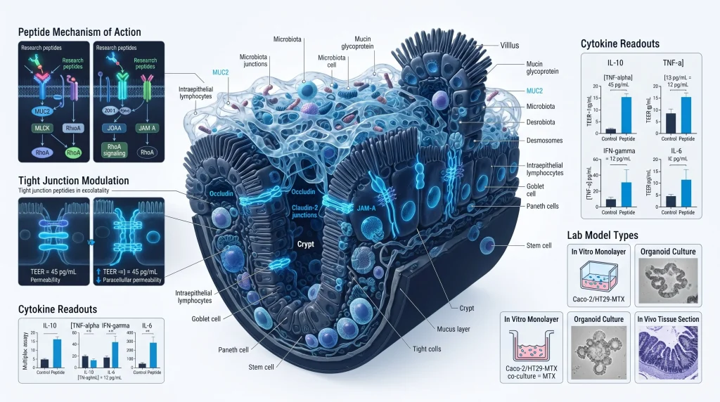
The gut mucosa is the inner lining of the small and large intestine. It is made of a single layer of gut cells. These cells form tight junctions that control what passes through the gut wall into the blood. Peptide gut mucosal biology research tests how specific compounds change tight junction protein levels, cytokine output, and epithelial repair in lab models.
Labs use gut cell cultures (such as Caco-2 and HT-29 cells) and rodent gut tissue to study mucosal biology. Cell cultures allow direct measurement of tight junction proteins. Rodent models add whole-gut context. Together, these approaches give a fuller picture of how peptides interact with mucosal biology at the cellular level.
How Does BPC-157 Act on Gut Mucosal Tissue?
BPC-157 (body protection compound-157) is a 15-amino-acid peptide first isolated from gastric juice. In gut mucosal lab models, BPC-157 adjusts cytokine balance. It lowers pro-inflammatory cytokines like IL-6 and TNF-alpha in gut tissue. It also affects nitric oxide (NO) output. NO controls blood flow and cell repair signals in gut epithelial tissue.
BPC-157 is also studied for its effects on tight junction proteins. Tight junctions are the links between gut cells. When tight junctions are stable, the gut barrier holds. Labs test BPC-157 in ulcer and mucosal injury models to see how it changes repair speed and cytokine output. According to PubMed (2019), BPC-157 adjusts cytokine levels in mucosal tissue models. Browse BPC-157 research vials for batch-tested supply.
What Role Does KPV Play in Gut Epithelial Models?
KPV (Lys-Pro-Val) is a tripeptide fragment of alpha-MSH (alpha-melanocyte stimulating hormone). In gut cell cultures, KPV blocks NF-kB signals. NF-kB (nuclear factor kappa-B) is the main path that drives pro-inflammatory cytokine output. When NF-kB is blocked, gut cells produce less IL-6, TNF-alpha, and IL-1beta.
KPV is studied in colitis and gut barrier models. In these assays, cytokine suppression and tight junction stability are the key readouts. According to PubMed (2015), KPV reduces cytokine output in gut cell models. Browse KPV research vials at Next Level Pharm for COA-verified lab supply.
How Is GHK-Cu Studied in Gut Repair Research?
GHK-Cu is a tripeptide (Gly-His-Lys) bound to a copper ion. Copper is needed for collagen cross-linking, the process that gives connective tissue its strength. In gut tissue models, GHK-Cu is studied for its role in collagen production and extracellular matrix (ECM) repair. The ECM is the scaffold below the gut cell layer. A strong ECM supports gut cell attachment and tissue integrity.
Labs apply GHK-Cu to gut tissue cultures to measure collagen output and ECM protein levels. They also test it in wound healing models where ECM repair is a key step. According to PubMed (2018), GHK-Cu affects collagen synthesis and growth factor output in tissue models. Browse GHK-Cu research vials for COA-verified supply.
What Is TB-500’s Role in Mucosal Repair Models?
TB-500 is a synthetic analog of thymosin beta-4 (TB4). TB4 is a natural peptide that promotes cell migration. In gut mucosal repair models, TB-500 promotes the movement of gut cells across wounds. Epithelial cell migration is a key step in closing mucosal gaps. Labs use TB-500 in scratch assay models to measure how fast gut cells cover a wound in a cell culture dish.
TB-500 also binds actin. Actin is the protein that forms the cell skeleton. When action is changed, cells move more freely. This makes TB-500 useful in gut mucosal migration assays where cell movement speed and direction are the key readouts. Labs often study TB-500 alongside cytokine-blocking peptides to see how cell migration and immune signals interact. Browse TB-500 research vials for lyophilized, COA-verified supply.
Research labs can shop research peptides for batch-tested, HPLC-verified gut mucosal peptide supply.
How Do Gut Mucosal Peptides Compare in Labs?
Labs choose gut mucosal peptides based on the specific biology they want to study. Cytokine suppression, ECM repair, and epithelial migration are distinct research targets.
| Peptide | Mechanism | Key Target | Main Lab Model |
| BPC-157 | Cytokine and NO balance | IL-6, TNF-alpha | Mucosal tissue, ulcer models |
| KPV | NF-kB block | IL-6, TNF-alpha | Intestinal cell cultures |
| GHK-Cu | Collagen and ECM | Collagen output | Gut tissue repair assays |
| TB-500 | Actin and cell migration | Epithelial repair | Scratch assay models |
Combining BPC-157 with KPV in one model lets labs study both cytokine balance and NF-kB suppression together. Adding GHK-Cu covers the ECM repair side of mucosal biology.
Frequently Asked Questions
What is peptide gut mucosal biology research?
Peptide gut mucosal biology research uses lab models to study how small protein chains interact with the gut lining. Labs track cytokine output, tight junction protein levels, gut cell movement, and collagen production. Common models include Caco-2 gut cell cultures and rodent gut tissue assays. BPC-157, KPV, GHK-Cu, and TB-500 are among the most studied compounds. All research is conducted for lab use only in preclinical settings.
What is the gut mucosal barrier and why do labs study it?
The gut mucosal barrier is the single layer of gut cells that lines the intestine. These cells form tight junctions. Tight junctions control what crosses the gut wall into the blood. When tight junctions are stable, the barrier holds. When tight junctions lose function, immune signals rise. Labs study how peptides change tight junction proteins and cytokine output to understand barrier biology in controlled preclinical models.
How does BPC-157 affect gut mucosal tissue in lab models?
BPC-157 is a 15-amino-acid peptide from gastric juice. In gut mucosal models, it adjusts cytokine balance and nitric oxide output. It also affects tight junction proteins. According to PubMed (2019), BPC-157 adjusts cytokine levels in mucosal tissue. Labs test it in ulcer and mucosal injury models to measure repair speed and cytokine changes. It is studied in cell cultures and rodent gut tissue under preclinical conditions.
What is KPV and how does it act in gut cell models?
KPV is a tripeptide (Lys-Pro-Val) derived from alpha-MSH. In gut cell cultures, KPV blocks NF-kB signal, the main driver of pro-inflammatory cytokine output. When NF-kB is blocked, cells produce less IL-6, TNF-alpha, and IL-1beta. According to PubMed (2015), KPV reduces cytokine output in gut cells. Labs use it in colitis and gut barrier models to measure cytokine suppression and tight junction stability.
What is GHK-Cu and how is it studied in gut repair models?
GHK-Cu is a tripeptide (Gly-His-Lys) bound to a copper ion. Copper is needed for collagen cross-linking in connective tissue. In gut tissue models, GHK-Cu is studied for collagen production and ECM (ECM) repair below the epithelial layer. Labs apply it to gut tissue cultures and measure collagen output and ECM protein levels. According to PubMed (2018), GHK-Cu affects collagen and growth factor output in tissue models.
What is TB-500 and how does it aid mucosal repair in lab models?
TB-500 is a synthetic analog of thymosin beta-4, a natural peptide that promotes cell migration. In gut mucosal repair models, TB-500 promotes gut cell movement across wound sites. It does this by binding actin, the protein that forms the cell skeleton. Faster cell movement speeds up the closing of mucosal gaps in scratch assay models. Labs study TB-500 in mucosal wound healing assays to understand how it changes epithelial migration speed and direction.
How do labs measure gut mucosal peptide effects?
Labs use several methods to measure mucosal peptide effects. ELISA measures cytokine levels in cell culture media. TEER (transepithelial electrical resistance) measures tight junction strength. Immunofluorescence imaging shows tight junction protein distribution in gut cell layers. Scratch assays measure gut cell movement speed. Collagen assays measure ECM output. Each method targets a different aspect of mucosal biology in controlled in vitro conditions.
What is TEER and why is it used in gut research?
TEER (transepithelial electrical resistance) is a measure of how well gut cells form a barrier. High TEER means tight junctions are intact and the barrier is strong. Low TEER means tight junctions are loose. Labs measure TEER in Caco-2 or T84 gut cell cultures. When a peptide is applied, TEER changes show whether the peptide supports or disrupts the gut mucosal barrier. TEER is a fast, non-destructive readout for gut barrier studies.
Can BPC-157 and KPV be combined in a gut mucosal study?
Combining BPC-157 and KPV in one gut model lets labs study cytokine balance (BPC-157) and NF-kB suppression (KPV) at the same time. This dual approach gives a fuller picture of how multiple immune signal paths interact in gut epithelial models. Labs can compare whether the two peptides have additive cytokine suppression effects. Multi-compound designs like this are common in gut mucosal research where cytokine, barrier, and repair signals all interact.
What purity is needed for gut mucosal peptide research?
Gut mucosal research requires high purity peptides because impurities can trigger cytokine responses in gut cell cultures. If a peptide batch contains residual solvents or synthesis byproducts, cell cultures may respond to those impurities rather than the peptide. Labs need peptide purity of 99% or higher for reliable cytokine and barrier assay results. HPLC testing confirms purity. Mass spectrometry confirms molecular identity. A COA links lot numbers to test results for each batch.
Summary
Peptide gut mucosal biology research tests how small protein chains interact with the gut lining in controlled lab models. BPC-157 adjusts cytokine balance and NO output in mucosal tissue. KPV blocks NF-kB to lower cytokine output in gut cell cultures. GHK-Cu supports collagen production and ECM repair in gut tissue assays. TB-500 promotes gut cell movement in mucosal wound healing models.
Each compound targets a distinct part of gut mucosal biology. Labs often combine cytokine-blocking peptides with ECM repair or migration compounds to study how these signals interact.
What Should You Do Next?
Researchers should define whether the gut model targets cytokines, tight junctions, extracellular matrix repair, or epithelial movement. Select the peptide and assay endpoints that match that target. Review the COA and record purity, molecular identity, and the lot number before each run. Researchers sourcing these lab peptides can shop research peptides with full COA and lot traceability.
People Also Read
- BPC-157 Research Peptide: Mucosal and Tissue Repair Studies
- KPV Research Peptide: Anti-Inflammatory Gut Epithelial Models
About the Author
Next Level Pharm Research Team
Alex M covers peer-reviewed findings in peptide science for Next Level Pharm, a US-based supplier of research-grade peptides verified to ≥99% purity via HPLC and mass spectrometry on every batch.
Disclaimer: For research purposes only. Not intended for human consumption. Next Level Pharm products are not intended for diagnostic, therapeutic, or medicinal use. This content does not constitute medical advice. Always consult a licensed healthcare professional before making any health-related decisions.