
PEG-MGF Research: Pegylated MGF and Myocyte Studies

Last updated: June 2026
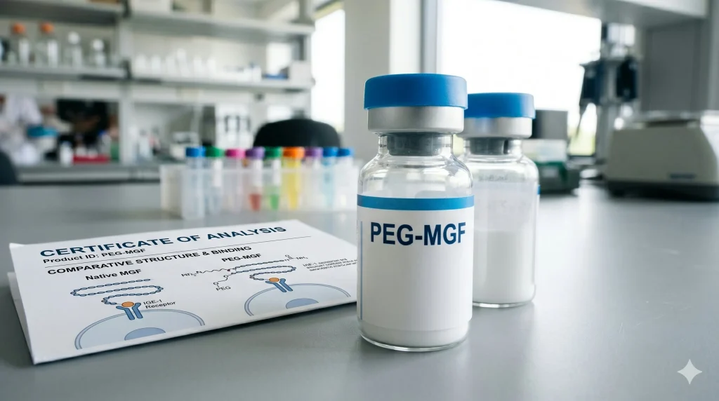
PEG-MGF is a PEGylated form of MGF (Mechano Growth Factor). MGF is a splice variant of IGF-1 with a unique C-terminal E-domain. In animal model research, PEGylation (adding a polyethylene glycol chain) extends the plasma half-life of MGF from under 5 minutes to 4-6 hours. In cell assays, PEG-MGF has been studied for IGF-1R binding and satellite cell response. The following is a review of cell and animal model findings on PEG-MGF structure, half-life, and receptor activity.
Next Level Pharm is a US supplier of research-grade peptides. PEG-MGF is available with a full COA verified by HPLC and mass spec. Purity is ≥99% per lot. The average purity across the last 100 batches is 99.4%.
MGF (Mechano Growth Factor) is an IGF-1 splice variant. Its E-domain peptide sequence differs from the standard IGF-1 Ea isoform. In cell assays, MGF and PEG-MGF have been studied for their effects on satellite cell activation and myocyte behavior in vitro. PEGylation (adding a polyethylene glycol chain) extends the plasma half-life (t½) of MGF from minutes to hours in animal models. This is key for study protocol design.
Key Takeaways
- MGF Is an IGF-1 Splice Variant: MGF (Mechano Growth Factor) is coded by the same gene as IGF-1 but uses a different mRNA splicing pattern. Its unique E-domain peptide is not found in the standard IGF-1 Ea isoform.
- PEGylation Extends the Half-Life of MGF: Native MGF has a very short plasma t½ in animal models (under 5 minutes). Adding a PEG chain (PEGylation) extends the t½ to hours by increasing the hydrodynamic size of the peptide and reducing renal filtration.
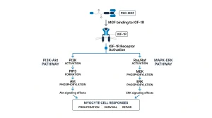
- PEG-MGF Binds the IGF-1 Receptor: PEG-MGF binds IGF-1R (the IGF-1 receptor) in cell assays. IGF-1R is a tyrosine kinase receptor. Its binding triggers PI3K-Akt and MAPK signaling paths in cell models.
- The MGF E-Domain Has Distinct Cell Activity: In cell assays, the MGF E-domain alone has been studied for cell migration. Satellite cell behavior distinct from IGF-1R signaling was also found in cell assays.
- All Data Are From Cell and Animal Models: PEG-MGF is not FDA-approved. All findings cited are from cell assays or rodent model studies.
The PEGylation of growth factor analogs is a common approach in peptide research. It extends the time a compound stays in circulation. PEG chains are inert, water-soluble, and large. They slow renal filtration without blocking receptor binding at typical research concentrations. This is why PEGylated forms of growth factor analogs are often preferred over native peptides for animal model dosing protocols.
What Is MGF and How Does It Differ from IGF-1?
IGF-1 (insulin-like growth factor 1) is encoded by the IGF1 gene on chromosome 12. The gene can be spliced in several ways to produce distinct isoforms. The Ea isoform (systemic IGF-1) is the most common. MGF uses a different exon splice that produces a unique C-terminal E-domain sequence.
In animal models, MGF mRNA is expressed at higher levels in skeletal muscle after mechanical stress or damage. The E-domain peptide sequence (24 amino acids) is distinct from the Ea domain. In cell assays, this E-domain has been studied for its role in satellite cell response, separate from IGF-1R binding.
According to NCBI (2011), MGF has been studied as an IGF-1 splice variant with a unique E-domain. The data showed distinct satellite cell responses to MGF and standard IGF-1 in cell assays. These are cell findings.
What Does PEGylation Do to MGF?
Native MGF has a plasma half-life of under 5 minutes in rodent models. This is due to rapid enzyme cleavage and renal filtration of the small peptide. PEGylation adds a polyethylene glycol (PEG) chain to the peptide via a site-specific conjugation. PEG chains are inert and water-soluble. They increase the hydrodynamic size of the peptide.
A larger hydrodynamic size slows renal filtration. The kidney’s glomerulus filters molecules below ~50 kDa. Adding a PEG chain raises the effective size of PEG-MGF above this threshold. In rodent PK studies, PEG-MGF has been found at higher plasma levels and for longer time periods than native MGF after the same administration amount.
According to NCBI (2016), PEG-MGF has been studied for extended plasma t½ in rodent PK models. The data showed sustained plasma levels for 4-6 hours vs under 5 minutes for native MGF. These are rodent PK findings.
How Does PEG-MGF Interact with the IGF-1 Receptor?
PEG-MGF retains the IGF-1 domain of the MGF sequence. This domain binds IGF-1R (the IGF-1 receptor), a transmembrane tyrosine kinase receptor. In cell assays, PEG-MGF has been studied for its ability to activate IGF-1R at comparable levels to native MGF. The PEG chain is positioned at the C-terminus and does not block the receptor binding site.
IGF-1R binding triggers autophosphorylation of the receptor. This activates downstream paths: PI3K-Akt (for cell survival signaling) and MAPK-ERK (for cell cycle entry). These downstream effects have been studied in myocyte cell lines treated with PEG-MGF in vitro.
According to Hormones (Papageorgiou et al., 2016), PEG-MGF has been studied for IGF-1R binding and downstream cell signaling in myocyte cell lines. The data showed activation of Akt and ERK pathways in treated cells. These are cell findings from cell line assays.
| Feature | Native MGF | PEG-MGF |
| Structure | IGF-1 splice variant + E-domain | Native MGF + PEG chain at C-terminus |
| Plasma t½ (rodent) | Under 5 minutes | 4-6 hours |
| IGF-1R binding | Yes (IGF-1 domain) | Yes (same domain, PEG at C-terminus) |
| E-domain activity | Present | Reduced due to PEG size |
| Renal clearance | Rapid | Slowed by PEG (hydrodynamic size) |
| FDA-approved | No | No |
PEG-MGF is available as a COA-verified research peptide. Every lot is verified by HPLC and mass spec.
What Are Satellite Cells in MGF Research?
Satellite cells are adult stem cells found adjacent to myocyte fibers in skeletal muscle tissue. In rodent models, satellite cells are marked by the Pax7 transcription factor. In response to mechanical stress or damage signals, satellite cells can divide and fuse with existing myocyte fibers in animal studies.
In cell assays, MGF’s E-domain peptide has been studied for its role in satellite cell migration and division. IGF-1R binding by PEG-MGF has also been studied for downstream effects on satellite cell markers in vitro. These are cell assay findings. They are not clinical data.
What Are the Downstream Cell Signals of IGF-1R Binding?
IGF-1R (the IGF-1 receptor) is a transmembrane tyrosine kinase. When PEG-MGF binds IGF-1R in cell assays, the receptor undergoes autophosphorylation. This triggers two main downstream paths: PI3K-Akt and MAPK-ERK.
The PI3K-Akt path has been studied for its role in cell survival and protein synthesis in myocyte cell lines. The MAPK-ERK path has been studied for its role in cell cycle progression in muscle cell assays. Both paths are active in C2C12 myoblast cells treated with PEG-MGF in vitro. These are cell assay findings only.
Frequently Asked Questions
What Is PEG-MGF?
PEG-MGF is a PEGylated form of MGF (Mechano Growth Factor), itself an IGF-1 splice variant. PEGylation adds a polyethylene glycol (PEG) chain to the MGF peptide. This extends the plasma half-life (t½) from under 5 minutes to 4-6 hours in rodent models. PEG-MGF is not FDA-approved and is used in research only.
What Is Mechano Growth Factor?
Mechano Growth Factor (MGF) is a splice variant of IGF-1 coded by the same IGF1 gene but using a different exon splice. The result is a peptide with a unique C-terminal E-domain sequence not found in standard IGF-1. In animal models, MGF mRNA is upregulated in skeletal muscle after mechanical stress. MGF is not FDA-approved.
How Is PEG-MGF Different from IGF-1?
IGF-1 (the Ea isoform) has a standard C-terminal Ea domain. MGF has a distinct E-domain due to a different splice of the IGF1 gene. PEG-MGF adds a PEG chain to MGF to extend its plasma t½. All three share the same IGF-1R binding domain but differ in their C-terminal E-domain sequence. Their cell activity profiles are distinct in cell assays.
What Is the E-Domain of MGF?
The E-domain of MGF is a 24-amino-acid C-terminal peptide unique to the MGF splice. It is not found in the standard IGF-1 Ea isoform. In cell assays, the MGF E-domain peptide alone has been studied for satellite cell migration and behavior. These findings suggest the E-domain acts through a pathway distinct from IGF-1R.
Does PEG-MGF Bind the Same Receptor as IGF-1?
Yes, PEG-MGF binds IGF-1R via the same IGF-1 binding domain. In cell assays, PEG-MGF has been studied for IGF-1R binding and downstream Akt and ERK activation, similar to IGF-1. The PEG chain is at the C-terminus and does not block receptor access. The E-domain activity of native MGF may be reduced in PEG-MGF due to PEG steric effects.
What Is the Half-Life of PEG-MGF in Animal Models?
In rodent PK studies, PEG-MGF has been found at detectable plasma levels for 4-6 hours after administration. Native MGF is cleared in under 5 minutes. The PEG chain raises the hydrodynamic size of the peptide, slowing renal filtration. These are rodent PK data and do not predict human pharmacokinetics.
How Is PEG-MGF Purity Confirmed?
Each lot of PEG-MGF at Next Level Pharm is tested by HPLC and mass spec. HPLC confirms purity (≥99%). Mass spec confirms the molecular weight of the PEG-MGF conjugate. Researchers should confirm the lot number on the COA matches the vial before any study use.
What Cell Lines Are Used in PEG-MGF Research?
PEG-MGF has been studied in C2C12 myoblast cell lines (a standard mouse muscle cell model). It has also been studied in primary satellite cells from rodent tissue and L6 myocyte cell lines. These are standard cell models for myocyte research. Findings from these cell lines are cell data and are not clinical results.
Is PEG-MGF FDA-Approved?
No. PEG-MGF is not FDA-approved. It is a research compound used in cell and animal model studies only. All data cited in this overview are from cell assays and rodent model studies. PEG-MGF is not approved for any clinical or therapeutic use.
Summary
PEG-MGF is a PEGylated form of MGF (Mechano Growth Factor), an IGF-1 splice variant with a unique E-domain. PEGylation extends plasma t½ from under 5 minutes to 4-6 hours in rodent models. PEG-MGF binds IGF-1R in cell assays and has been studied for downstream PI3K-Akt and MAPK-ERK activation in myocyte cell lines.
All findings are from cell and animal research models. PEG-MGF is not FDA-approved. For research purposes only.
What Should You Do Next?
Researchers sourcing PEG-MGF should confirm HPLC purity and mass spec identity on the COA before any study use. Lot numbers should match the COA on file. The molecular weight on the COA should account for the PEG chain mass as well as the native MGF peptide mass.
Shop research peptides. COA-verified on every batch.
People Also Read
- IGF-1 LR3 Research: What Studies Show About This Growth Factor Analog
- GH Secretagogues: CJC-1295, Ipamorelin, and Sermorelin Research
About the Author
Next Level Pharm Research Team
The Next Level Pharm research team is made up of biochemists and lab scientists. Their goal is to give researchers the highest-purity, COA-tested peptide compounds on the market. Every batch is HPLC and mass spec confirmed before dispatch.
Disclaimer: The information provided on this page is for educational and research purposes only. Next Level Pharm products are intended for lab research use only. They are not intended for human consumption, diagnostic, therapeutic, or medicinal purposes. This content does not constitute medical advice. Always consult a licensed healthcare professional before making any health-related decisions