
Nrf2 ARE Pathway Peptide Research: Antioxidant Studies

Last updated: October 2026
A Nrf2 ARE antioxidant pathway is a cellular defense system that controls genes for antioxidant and detox enzymes. Nrf2 stands for nuclear factor erythroid 2-related factor 2. ARE stands for antioxidant response element. According to PubMed (2003), Nrf2 is a key transcription factor that drives expression of over 200 protective genes. Peptide researchers study how specific peptides change this pathway in cell and tissue models.
Next Level Pharm supplies research-grade peptides at 99.4% average purity. Each batch is tested by HPLC and mass spec before dispatch. A COA ships with every vial. Lot numbers are traceable online. Peptides arrive as lyophilized powder and are stable at room temp during shipping. Third-party testing is part of every batch run, and results appear on the COA alongside in-house data.
Nrf2 ARE pathway peptide research studies how lab compounds activate antioxidant gene networks in cell models.
Nrf2 research covers three core areas: pathway activation, reporter assays, and Keap1 binding studies. Each area relies on high-purity peptide material for clean data. The Key Takeaways below outline the main concepts.
Key Takeaways
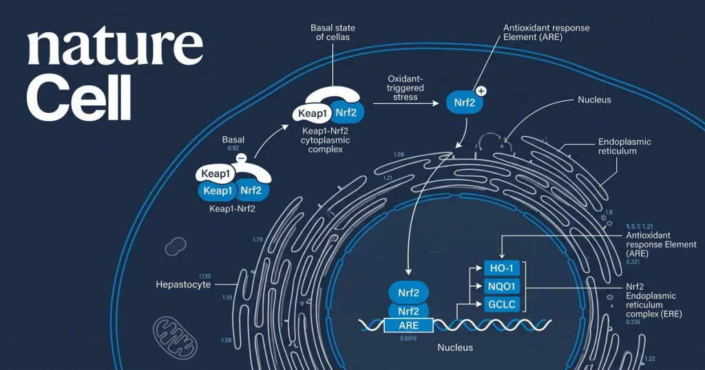
- Nrf2 Activation: Nrf2 is kept in the cytoplasm by Keap1 and moves to the nucleus when the Keap1 bond breaks.
- ARE Binding: Once in the nucleus, Nrf2 binds ARE sequences and turns on genes like HO-1 and NQO1.
- Peptide Modulators: Some peptides activate Nrf2 by modifying Keap1 cysteine residues or by direct protein binding.
- Reporter Assays: ARE-luciferase reporters in HEK293 or HepG2 cells give a fast, numeric readout of Nrf2 activity.
- Keap1-Nrf2 Interface: The binding interface is a main target for peptide probe design in pathway studies.
- Purity Standard: Peptide impurities can trigger false-positive Nrf2 activation, so 99.4% purity is the baseline.
Beyond receptor binding, Nrf2 pathway research links to mitochondrial health and redox signaling. Lab studies in each area depend on COA-verified research peptides for clean results.
What Is the Nrf2 ARE Antioxidant Pathway?
Nrf2 is a transcription factor. It controls a large set of antioxidant and detox genes. Under normal conditions, Keap1 holds Nrf2 in the cytoplasm. Keap1 marks Nrf2 for rapid breakdown. When the cell faces oxidative stress, Keap1 cysteine residues get modified. This releases Nrf2 from the complex. Nrf2 then moves to the nucleus and binds ARE sequences. This starts expression of protective genes, including HO-1, NQO1, and GCLC.
According to NIH (2013), the Nrf2-Keap1 system acts as the main cellular sensor for reactive oxygen species. The Keap1 protein has 27 cysteine residues. Some of these are highly reactive sensors. Peptides that signal this pathway can trigger Nrf2 release without causing cell damage. This makes them useful probes for pathway studies in lab models. Next Level Pharm supplies research-grade peptides for in vitro Nrf2 work.
How Do Peptides Activate Nrf2 Nuclear Entry?
Peptides activate Nrf2 by two main routes. The first route is indirect. A peptide modifies Keap1 cysteine residues. This loosens the Keap1-Nrf2 complex and frees Nrf2. The second route is direct. A peptide binds the Kelch domain of Keap1. This blocks the Nrf2 docking site. Both routes cause Nrf2 to accumulate in the nucleus.
According to Frontiers in Pharmacology (2022), short peptides modeled on the ETGE binding motif can block Keap1 and trigger Nrf2 nuclear entry. This points to the ETGE region as a viable peptide probe target. Knowing which route a test peptide takes helps researchers read ARE reporter results. A peptide that acts via cysteine changes gives a broad redox signal. A peptide that acts via direct binding gives a cleaner, target-specific readout.
What Assays Measure ARE Reporter Activity In Vitro?
The main assay for Nrf2 activity is the ARE-luciferase reporter. A plasmid with ARE sequences drives a luciferase gene in the test cell. When Nrf2 activates, luciferase output rises. A plate reader measures this as fold-change over vehicle control. The assay runs in 96-well format and scales well for screening many compounds.
According to PubMed (2024), ARE-reporter assays in HepG2 and HEK293 cells are the standard tool for ranking Nrf2 activator potency. Secondary assays confirm downstream gene expression. qPCR measures HO-1 and NQO1 mRNA. Western blotting of nuclear Nrf2 validates the reporter signal. Together, these three methods give a complete picture of Nrf2 activation. Peptide purity is key here. A 5% impurity can produce false-positive ARE induction from off-target chemistry.
Browse antioxidant research peptides at Next Level Pharm for COA-verified research peptides used in Nrf2 pathway studies.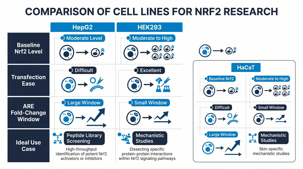
How Does Nrf2 Interact with Keap1 in Signaling Studies?
The Nrf2-Keap1 interface has two binding motifs on Nrf2: ETGE and DLG. Both motifs dock to the Kelch domain of Keap1. The ETGE motif binds with higher affinity than DLG. Under resting conditions, both motifs bind and lock Nrf2 for breakdown. Stress breaks the DLG-Kelch bond first. This lets Nrf2 escape while ETGE stays attached. This hinge-latch model explains partial Nrf2 activation.
According to NCBI PMC (2012), co-IP and surface plasmon resonance, or SPR, are the main methods to measure Keap1-Nrf2 binding in vitro. SPR gives on-rate, off-rate, and KD values for peptide probes at the Kelch domain. This data helps rank probe affinity. Peptides such as SS-31 also intersect with redox and mitochondrial pathways, which can overlap with Nrf2 studies in some lab designs.
What Cell Lines Are Used for Nrf2 Pathway Research?
Three cell lines dominate Nrf2 pathway research. HepG2 cells are human liver cells. They have high baseline Nrf2 and a wide fold-change window. HEK293 cells are human kidney cells. They are used for transfection-based ARE-luciferase assays. HaCaT cells are human skin keratinocytes. They are used in Nrf2 studies of barrier and UV stress models. Each line has a different baseline Nrf2 level, which affects fold-change results for test peptides.
According to Frontiers in Cell and Developmental Biology (2021), HepG2 cells show a wider fold-change range and are preferred for initial screening. HEK293 cells are easy to transfect and give stable reporter signal across batches. Researchers often test in both lines to confirm activity is not cell-type specific. Research peptides such as Semax have been studied alongside Nrf2 pathway markers in oxidative stress cell models.
How Are Nrf2 Pathway Peptides Verified for Research Use?
Research-grade Nrf2 pathway peptides are tested by HPLC and ESI-MS before dispatch. HPLC confirms the purity profile by measuring the main peak area against the total area. ESI-MS verifies the correct molecular mass and screens for truncated forms or oxidized residues. Both data sets appear in full on the COA for each lot.
The USP states that research peptide identity testing should include HPLC retention time and mass ion data. These standards are applied to every product in the catalog. The average purity across the last 100 batches is 99.4%. Each lot gets a unique traceable lot number. Researchers can check lot numbers online before or at the time of order. Lyophilized peptides for Nrf2 studies are stable at room temperature during transit. Store at minus 20 C on receipt for long-term stability.
Frequently Asked Questions
What is the Nrf2 ARE antioxidant pathway?
The Nrf2 ARE pathway is a cellular defense system. It controls genes for antioxidant and detox enzymes. Nrf2 is normally held in the cytoplasm by Keap1 and broken down rapidly. Oxidative stress or reactive compounds release Nrf2 from Keap1. Nrf2 then moves to the nucleus and binds ARE sequences. This activates protective genes like HO-1 and NQO1 to reduce oxidative load in the cell.
How do peptides activate Nrf2 nuclear entry?
Peptides activate Nrf2 by two routes. The first route is indirect: a peptide modifies Keap1 cysteine residues and loosens the Keap1-Nrf2 bond. The second route is direct: a peptide binds the Kelch domain of Keap1 and blocks the Nrf2 docking site. Both routes lead to Nrf2 nuclear entry. The direct route gives a cleaner, target-specific readout in assays. Lab studies track nuclear entry by fractionation or by GFP-tagged Nrf2 imaging.
What assays measure ARE reporter activity in vitro?
The ARE-luciferase reporter assay is the standard tool for Nrf2 activity in lab work. A plasmid with ARE sequences drives a luciferase gene in the test cell. When Nrf2 activates, luciferase signal rises and is read as fold-change over vehicle. Secondary assays confirm the signal: qPCR measures HO-1 and NQO1 mRNA. Western blotting measures nuclear Nrf2 protein. All three methods together give a complete picture of Nrf2 activation in the model.
How does Nrf2 interact with Keap1 in signaling studies?
Nrf2 carries two binding motifs, ETGE and DLG, that both dock to the Kelch domain of Keap1. The ETGE motif binds with higher affinity than DLG. Under stress, the DLG-Kelch bond breaks first, which lets Nrf2 start to build up while ETGE stays attached. This hinge-latch model explains partial Nrf2 activation. SPR and co-IP are the main in vitro methods to measure this binding. They give KD and kinetic values for peptide probes at the interface.
What cell lines are used for Nrf2 pathway research?
HepG2, HEK293, and HaCaT are the three main cell lines for Nrf2 work. HepG2 cells are human liver cells with high baseline Nrf2 and a wide fold-change window. They are the first choice for screening peptide activators. HEK293 cells are easy to transfect and give stable ARE reporter signal across batches. HaCaT cells are skin keratinocytes used in UV and barrier stress models. Researchers often test in two lines to confirm results are not cell-type specific.
What is the role of HO-1 in Nrf2 pathway studies?
HO-1, or heme oxygenase 1, is one of the main target genes activated by Nrf2. Its promoter has ARE sequences that Nrf2 binds directly. In lab studies, HO-1 mRNA or protein level is used as a readout to confirm Nrf2 activity. A rise in HO-1 after peptide treatment, measured by qPCR or western blot, supports ARE pathway activation. HO-1 is a functional marker because its signal is strong and easy to measure in most lab cell lines.
Why does peptide purity matter in Nrf2 assays?
Peptide purity is critical in Nrf2 assays because impurities can trigger false-positive ARE activation. Truncated peptide fragments or oxidized forms can act as reactive compounds. They modify Keap1 cysteines and release Nrf2 without a specific ligand effect. A 5% impurity level can produce an ARE fold-change signal that looks like real pathway activation. HPLC-verified 99.4% purity material is the standard for clean, readable Nrf2 assay data in lab settings.
How are Keap1-Nrf2 binding studies run in vitro?
Keap1-Nrf2 binding is studied by SPR, AlphaScreen, or co-IP. SPR uses a sensor chip coated with recombinant Keap1 Kelch domain. Test peptides are flowed over the chip and on-rate and off-rate constants are measured. These give a KD value for each peptide. AlphaScreen uses bead-based proximity detection and runs in 384-well format for higher output. Co-IP confirms binding in a cell context and is run as a secondary check after biophysical methods.
What is the ETGE motif in Nrf2 research?
The ETGE motif is a short amino acid sequence on Nrf2 that docks to the Kelch domain of Keap1. It binds with high affinity, which makes it the main anchor of the Nrf2-Keap1 complex. Short synthetic peptides based on the ETGE sequence can compete with full-length Nrf2 for Keap1 binding in vitro. These peptides are used as tool compounds to study the Keap1-Nrf2 interface. They help researchers find which Kelch residues drive the interaction.
How do researchers confirm Nrf2 nuclear entry?
Nuclear entry is confirmed by one or more of three methods. The first is fractionation, which splits the cell into nuclear and cytoplasmic portions and measures Nrf2 by western blot in each. The second is staining with an anti-Nrf2 antibody, which shows Nrf2 location in fixed cells. The third is a GFP-Nrf2 fusion, which lets researchers track Nrf2 movement in live cells by fluorescence. All three methods need a validated anti-Nrf2 antibody or a confirmed GFP fusion construct.
Summary
Nrf2 ARE antioxidant pathway research studies how peptides and other lab compounds activate the Keap1-Nrf2 system to drive antioxidant gene expression. The pathway centers on Nrf2 nuclear entry and ARE binding after Keap1 is disrupted. ARE-luciferase reporters in HepG2 and HEK293 cells are the main assay tools. HO-1 qPCR and nuclear Nrf2 western blot serve as secondary confirmation.
Peptide purity is a key variable in this field. Oxidized forms or truncated fragments can trigger false-positive ARE activation through non-specific chemistry. Research-grade peptides at 99.4% purity, confirmed by HPLC and mass spec, are the standard for clean, reproducible Nrf2 assay data.
What Should You Do Next?
- Select the ARE reporter cell line that fits your study: HepG2 for a wider fold-change range or HEK293 for stable transfection.
- Confirm test peptide purity by HPLC before running the ARE-luciferase assay.
- Validate Nrf2 activation with at least one secondary readout: HO-1 qPCR or nuclear fractionation western blot.
Shop research peptides for COA-verified research peptides with HPLC and mass spec data on every lot.
People Also Read
- SS-31 Peptide Research: Mitochondria and Cardiolipin Binding Studies
- Humanin Peptide Research: Mitochondrial Protection and Cytoprotection Studies
About the Author
Next Level Pharm Research Team
The Next Level Pharm research team is composed of biochemists and laboratory scientists dedicated to providing researchers with the highest-purity, COA-verified research peptides available. Every batch is HPLC and mass spec verified before dispatch.
Disclaimer: The information provided on this page is for educational and research purposes only. Next Level Pharm’s products are intended for laboratory research use only. They are not intended for human consumption, diagnostic, therapeutic, or medicinal purposes. This content does not constitute medical advice. Always consult a licensed healthcare professional before making any health-related decisions.