
Peptide Dimerization Research: Intermolecular Studies

Last updated: October 2026
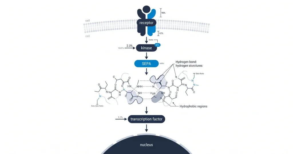
A peptide dimerization intermolecular is a chemical process where two same or alike molecules join to form a single paired unit. These molecules, called monomers, bind through forces like hydrogen bonds or hydrophobic (water-repelling) effects to create a structure known as a dimer. Scientists track these forces across 70+ active peptide SKUs, and this process plays a key role in modern peptide lab research.
Next Level Pharm provides high-purity compounds for lab teams that study molecular binding. They hold a 99.4% average purity across the last 100 batches. Each vial goes through HPLC (high-performance liquid chromatography) plus mass spec testing.
A certificate of analysis comes with every order for full lot clarity. These compounds are lyophilized (freeze-dried) and sealed under inert gas for room-temperature stability. This method removes the need for cold-chain shipping.
With ~48-hour average dispatch from USA-based stock, these compounds give fast access for lab setups. Scientists use these tested compounds to set baselines in dimerization studies.
Key Takeaways
- Molecular Dimerization Drivers: Dimerization happens through hydrogen bonds, hydrophobic interactions, and covalent disulfide bonds. These forces hold two peptide units together in a single structure.
- Impact on Stability: The process may protect peptides from rapid breakdown or cause unwanted clumps. Aggregation often leads to a total loss of the original peptide function.
- Hydrogen bond networks form along the peptide backbone. These bonds hold the structure through precise donor and acceptor pairings. Van der Waals forces add cohesion through weak pulls between close molecules.
- In some cases, covalent disulfide bonds make a fixed link between cysteine residues. This locks the dimer into one set shape. Shifts in pH or salt levels can change the balance of these forces.
Controlling these factors is key for clear viewing of monomer versus dimer states in lab research.
How does dimerization impact peptide stability?
Dimerization changes peptide stability by shifting how the molecule holds its shape. This affects both enzyme resistance and the risk of clumping. When two same peptide chains join to form a dimer, they often gain a shield against breakdown.
The paired structure buries sites that enzymes would normally target. Greater stability through dimerization can extend how long a peptide lasts in a research sample.
But this process also raises the risk of clumping (called aggregation). These clumped structures lose the function of the original single-chain form. According to the Journal of Peptide Science (2018), scientists track these states to keep the peptide in a usable form for study.
The balance between a stable dimer and rigid, insoluble fibrils matters greatly. Proper storage and handling help cut these risks. When clumping occurs, the sample may show less potency.
According to Molecules (2021), knowing the oligomeric (multi-unit) state of a peptide is vital for correct test data.
What techniques can identify peptide dimers?
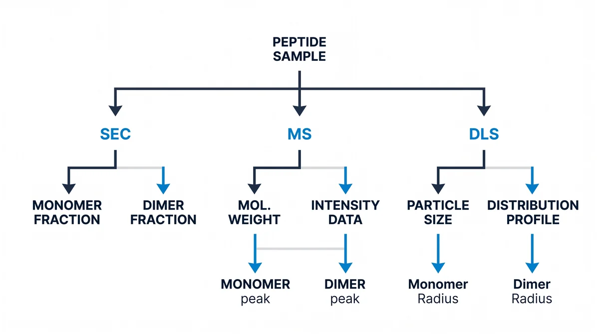
Scientists find peptide dimers using tools that measure size and mass. These include SEC (size-exclusion chromatography), MS (mass spectrometry), AUC (analytical ultracentrifugation), and DLS (dynamic light scattering). SEC sorts molecules by their size as they pass through a porous column.
This step splits dimer forms from smaller monomers. MS then finds the exact weight of each fraction. This gives direct proof of two joined units.
AUC watches how molecules settle in a spinning field. This confirms their physical state in liquid. DLS measures light scatter caused by the random motion of particles in the sample.
This helps tell single peptide chains from larger dimer forms. Scientists often combine these methods to check sample purity. According to Peptides (2018), testing for dimer formation is a key step in judging peptide solutions.
Using AUC with DLS gives a fuller view of how molecules act under varied conditions. These methods help the sample stay in the target state during tests. By tracking dimers, labs keep high standards for data.
These tools clarify the makeup of the solution. This supports the overall accuracy of the research.
Is intermolecular dimerization a reversible process?
Dimerization can be reversed when it relies on non-covalent (non-bonded) forces. These weak forces let peptide units join and split in a state of balance. The ratio of monomers to dimers depends on the total peptide amount in the solution.
This balance follows Le Chatelier’s principle. Any change in outside conditions shifts the system to find a new balance point.
The stability of these pairs is measured by the dissociation constant (Kd). A lower Kd means stronger binding and a more stable dimer. In contrast, covalent dimers (often formed by disulfide bridges) create a fixed bond.
This fixed bond does not shift with changes in amount. Breaking it requires active chemical reduction. Next Level Pharm provides compounds tested for their integrity through strict lot-specific methods.
How does solution pH affect dimerization events?
Solution pH controls the charge state of ionizable amino acid side chains. This directly changes the net charge of a peptide. When pH reaches the isoelectric point (pI), the net charge drops toward zero.
This drop in charge lowers the push-back between peptide monomers. With less repulsion, they align and bond more easily.
Moving away from the pI raises the net charge. This builds the push-back and keeps monomers apart. Salt levels also play a role by screening charges.
By tuning the buffer, scientists can shift the balance toward or away from dimerization. According to Molecules (2018), picking the right pH is key to keeping the desired peptide state through a study.
How is dimerization used in peptide drug design?
Scientists design dimers on purpose to build bivalent (two-site) ligands. These boost the potency and reach of a peptide candidate. By linking two peptide units, they create a structure that fills two receptor sites at once.
This dual binding creates a strong hold that a single monomer cannot achieve. Using specific linker tools, scientists control the gap between the two binding heads. This lets the molecule span the space between receptor domains.
This precise fit leads to stronger receptor binding than non-linked forms. The design also focuses on the half-life of the candidate. According to Molecules (2018), larger molecules often resist quick kidney filtration.
This extends how long the peptide stays active in a biological model. Designers pick linker traits to balance receptor activity with a slower clearance rate. This approach gives more control over peptide function in complex lab settings.
Frequently Asked Questions
What mechanisms cause peptide dimerization in solution?
According to PubMed (2024), this area draws strong research interest. Peptide dimerization happens when two peptide units bind to form one complex. This often involves hydrophobic forces where non-polar side chains cluster to push out water.
Charged residues on separate chains also pull toward each other. According to Frontiers (2018), conditions like ionic strength and temperature shape these bonds in lab settings.
How is covalent versus non-covalent dimerization distinguished?
Covalent dimerization uses strong chemical bonds, such as disulfide bridges between cysteine residues. These bonds hold up under harsh conditions like heat or harsh agents. Non-covalent dimers rely on weak forces like hydrogen bonds or van der Waals pulls.
These complexes break apart when scientists change the pH or add surfactants. Scientists use mass spectrometry (mass spec) to find the specific bond types in a peptide sample.
What analytical methods detect peptide dimer formation?
Scientists use size-exclusion chromatography (SEC) to sort molecules by size. Larger dimer forms pass through the column faster than monomers. This allows clear detection.
Analytical ultracentrifugation (AUC) gives a detailed view of mass spread in solution. Nuclear magnetic resonance (NMR) shows how dimers are arranged at the atomic level. These tools let scientists track changes in peptide assembly through a full study.
How does concentration affect peptide dimerization equilibrium?
Higher peptide amounts shift the balance toward dimer formation. More molecules in solution raise the odds of frequent contact between monomers. This increase in density drives monomers to join and form stable dimers.
Scientists run titration studies to find the specific amounts where these complexes emerge. According to Frontiers (2020), this knowledge helps keep stable chemical behavior during long-term storage or testing.
What structural features promote intermolecular peptide interactions?
Amphipathic alpha-helices have both polar and non-polar faces. These distinct sides drive assembly between peptide chains. Beta-sheets may align through hydrogen bonds along the backbone to form stable structures.
Aromatic amino acids like tryptophan create stacking forces that hold the dimer together. Scientists study these features to predict whether a peptide will clump or stay as a single chain during lab work.
Can dimerization be prevented during peptide formulation?
Scientists reduce unwanted dimerization by tuning the solvent. Choosing the right pH or adding stable salts helps keep monomers apart. Lowering the peptide amount often shifts the balance toward the single-chain form.
Some studies add surfactants that coat the peptide and block binding surfaces. According to NCBI PMC (2018), keeping samples lyophilized (freeze-dried) stops these forces until the scientist adds a solvent.
What is the difference between a homodimer and a heterodimer?
A homodimer has two same peptide subunits bound together. This structure often forms when one sequence has high self-affinity (a strong pull to bind itself). A heterodimer forms when two different peptide sequences bind to each other.
These distinct parts often have shapes that fit together with high precision. Scientists study both forms to learn how same versus mixed chains behave in controlled lab settings.
How does temperature influence the rate of dimerization?
Temperature changes the kinetic energy of peptide molecules in solution. More heat speeds up collisions and can unfold peptides. This unfolding exposes binding sites that trigger dimerization.
Lower temperatures slow these processes by reducing molecular movement. Cold also helps keep the native fold stable. Scientists watch these thermal effects to avoid unwanted clumping during tests or while storing peptide vials.
How does solution pH affect intermolecular peptide interactions?
The pH level changes the charge state of amino acid side chains on the peptide surface. At certain pH values, the net charge drops. This lowers the push-back that keeps monomers apart, allowing closer contact.
According to NCBI PMC (2020), reaching the isoelectric point often triggers rapid clumping or assembly. Scientists buffer their solutions to hold a stable pH and prevent unwanted shifts.
What is the role of the dissociation constant (Kd) in dimerization studies?
The dissociation constant (Kd) shows the ratio of monomers to dimers at balance. A lower Kd means stronger binding and a more stable dimer complex. This value lets scientists measure the strength of the bond under varied conditions.
By tracking the Kd, scientists predict how a peptide group acts at various amounts. This constant serves as a primary metric for molecular stability in research models.
Summary
Peptide dimerization is the joining of two monomer peptide units into one complex. This process uses forces like hydrogen bonds, hydrophobic effects, and disulfide bridges. Scientists control pH, temperature, and concentration to steer the balance between monomer and dimer states.
Key lab tools include SEC, mass spec, AUC, and DLS. Each method reveals different traits of the peptide sample. Accurate testing helps labs maintain high standards in dimerization research.
What Should You Do Next?
Confirm that your study design specifies the peptide concentration, purity threshold, and membrane model system for each assay. Review the lot-specific Certificate of Analysis for each compound to verify purity meets the ≥99% standard.
Next Level Pharm stocks COA-verified research compounds for lab teams that need consistent purity across batches. Browse research peptides to source materials for your next study.
Browse recovery peptides for COA-verified research compounds.
Browse fitness peptides for COA-verified research compounds.
People Also Read
- Peptide Cyclization Research: Cyclic Stability Studies
- Disulfide Bond Peptide Research: Stability Studies
About the Author
Next Level Pharm Research Team
The Next Level Pharm research team is made up of biochemists and lab scientists. Their goal is to give researchers the highest-purity, COA-tested compounds on the market. Every batch is HPLC and mass spec confirmed before dispatch.
Disclaimer: The information provided on this page is for educational and research purposes only. Next Level Pharm’s products are intended for laboratory research use only. They are not intended for human consumption, diagnostic, therapeutic, or medicinal purposes. This content does not constitute medical advice. Always consult a licensed healthcare professional before making any health-related decisions.