
Galanin Peptide Research: GalR Neuropeptide Receptor

Last updated: October 2026
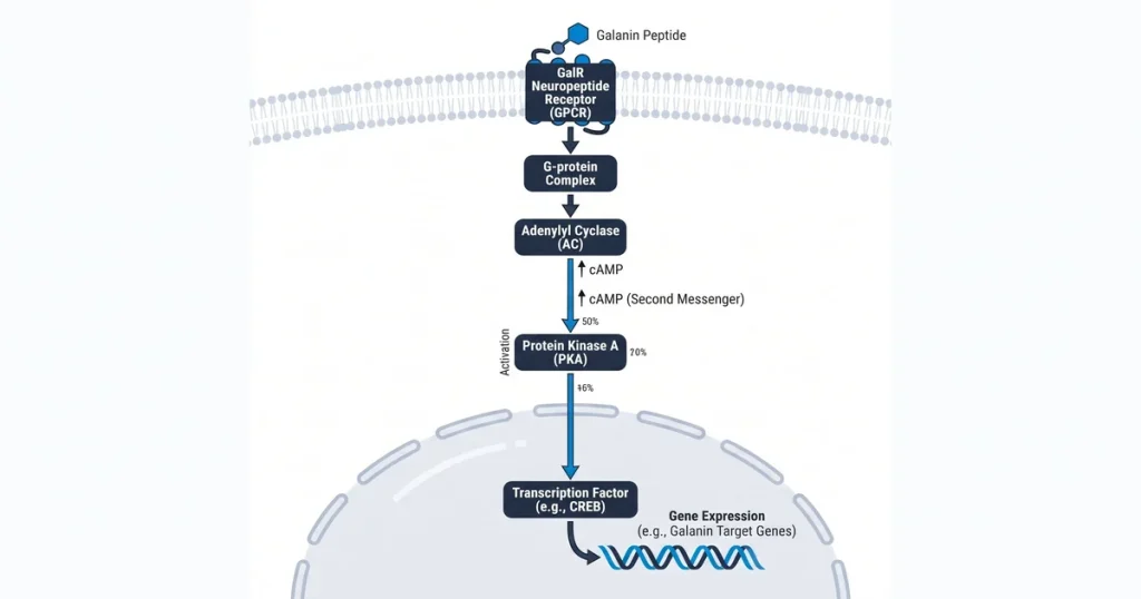
A galanin GalR neuropeptide receptor peptide research study is a lab exam of a short peptide found in the central and peripheral nervous systems. This peptide binds three GPCR subtypes: GalR1, GalR2, and GalR3. Each subtype fires a distinct cell path, and mapping these paths helps labs study how neuropeptide signals shape neural circuits in vitro.
Next Level Pharm supplies COA-verified galanin compounds at 99.4% purity across the last 100 batches. Every vial is HPLC and mass spec verified before dispatch. The peptide ships lyophilized and stable at room temperature, with a full COA and lot number on each order.
Receptor isolation studies require tight purity control. Small impurities can mask a single GalR subtype’s effect. Clean compounds are needed to draw clear conclusions from any signaling assay.
Key Takeaways
- Peptide Structure: Galanin is a 29-30 amino acid peptide. It is conserved across species, making it useful in diverse lab models.
- Three Receptor Subtypes: GalR1, GalR2, and GalR3 are each GPCRs. Each has a distinct G protein partner and a separate cell path.
- Signaling Divergence: GalR1 and GalR3 link to Gi proteins and lower cAMP. GalR2 links to Gq proteins and raises cell calcium.
- Research Tools: Galnon is a synthetic agonist that mimics galanin. M35 is an antagonist that blocks receptor binding in assays.
- Future Focus: Scientists are building selective ligands to isolate each receptor. This cuts off-target effects in neurological models.
The sections below cover each receptor mechanism in detail. They also describe the tools researchers use to isolate specific signaling paths.
What is the galanin neuropeptide?
Galanin is a neuro-modulatory peptide of 29-30 amino acids. It acts as a messenger in both the CNS and PNS. It was found by Tatemoto and Mutt in 1983 from porcine extracts.
According to PubMed (2007), galanin shapes how neurons fire across synaptic gaps. Its wide spread links it to mood, metabolism, and endocrine function. Because the peptide is conserved across species, it can be used in many lab models.
This broad spread gives it wide utility in neuroscience programs. Each study adds to the map of how amino acids regulate neural signals.
What are the three GalR receptor subtypes?
GalR1, GalR2, and GalR3 each bind galanin on the cell surface. All three are GPCRs. Each sits in the cell membrane and links an outside signal to an internal cell response.
Key differences across the three subtypes:
- GalR1: Found mainly in the brain and spinal cord. Links to Gi proteins, which lower cAMP inside the cell.
- GalR2: Found in both the CNS and peripheral tissues. Links to Gq proteins, which raise calcium and fire PKC.
- GalR3: Found in the hypothalamus and brainstem. Also links to Gi proteins and lowers cAMP, like GalR1.
According to the Journal of Peptide Science (2015), each subtype shows distinct tissue patterns. Researchers use transfected cell lines to isolate each subtype for clean pathway analysis.
How does galanin affect feeding behavior studies?
Galanin acts in the hypothalamus to shift hunger signals toward fat intake. It works inside the PVN (paraventricular nucleus). There it adjusts circuits that control satiety and food choice.
Galanin also works with NPY (neuropeptide Y) in the PVN. According to Peptides (2018), NPY drives general hunger while galanin steers toward fat intake. This two-peptide system creates a layered control over feeding signals.
Researchers study these paths in lab models to map hypothalamic energy circuits. The ratio of these two peptides shapes the type and strength of hunger signals.
Labs running galanin receptor studies can browse the recovery peptide catalog at Next Level Pharm.
What is galanin’s role in mood circuit studies?
Galanin shows region-dependent effects on mood-related neural circuits. In the locus coeruleus, it acts as an inhibitory signal. It slows stress-linked neuron firing in that region.
In the amygdala, GalR2 and GalR3 activation links to stress-response signaling changes in lab models. This dual behavior depends on local receptor density and peptide levels. When galanin slows the locus coeruleus, it can reduce stress arousal signals.
According to NCBI (2024), selective tools are now being used to map which receptor drives each effect. Labs studying stress-response models can browse brain peptides for related compounds.
How do GalR agonists and antagonists work?
An agonist binds to a GalR receptor and activates it. An antagonist binds to the same site but blocks activation. Both tools map receptor function in cell assays.
Key synthetic tools in GalR studies:
- Galnon: A non-peptide agonist that can enter cells. It simulates receptor activation in cell-based assays.
- M35: A peptide antagonist that blocks galanin from binding. It shows what happens when the path is cut off.
- Galanin (1-16): A short form that binds GalR1 and GalR2 but not GalR3. It helps distinguish which subtype drives a response.
Each tool gives researchers a different view of receptor function. Comparing agonist vs. antagonist results links a response to one subtype.
What are future galanin research directions?
Future work focuses on building ligands that target only one GalR subtype. General agonists fire all three subtypes at once. This makes it hard to isolate one receptor’s role.
Active areas include seizure threshold, neuronal survival, and mood circuit research. GalR1 agonists are studied for effects on seizure thresholds. GalR2 activation is tracked for links to neuron survival signals.
Designing selective ligands is hard because the three binding pockets share similar shapes. Progress here will allow finer mapping of how each subtype affects neural and metabolic signaling.
| Feature | GalR1 | GalR2 | GalR3 |
|---|---|---|---|
| G Protein | Gi/o | Gq/11 | Gi/o |
| Effect on cAMP | Lowers | No direct effect | Lowers |
| Calcium Release | No | Yes | No |
| Primary Location | Brain and spinal cord | CNS and periphery | Hypothalamus and brainstem |
| Key Study Use | Seizure models | Neuroprotection | Metabolic signaling |
Frequently Asked Questions
What are the GalR1 and GalR2 galanin receptor subtypes?
GalR1 and GalR2 are two of the three GPCR subtypes that bind galanin. GalR1 sits mainly in the brain and spinal cord. It links to Gi proteins and lowers cAMP in the cell.
GalR2 is found in the CNS and body tissues. It links to Gq and raises cell calcium instead. These two subtypes drive different cell responses, so researchers study them separately.
How does galanin bind to its receptor subtypes?
Galanin binds each GPCR subtype via contacts between its N-terminal chain and the receptor’s membrane domain. Each subtype has a slightly different binding pocket. Small changes in peptide structure can shift binding from one subtype to another.
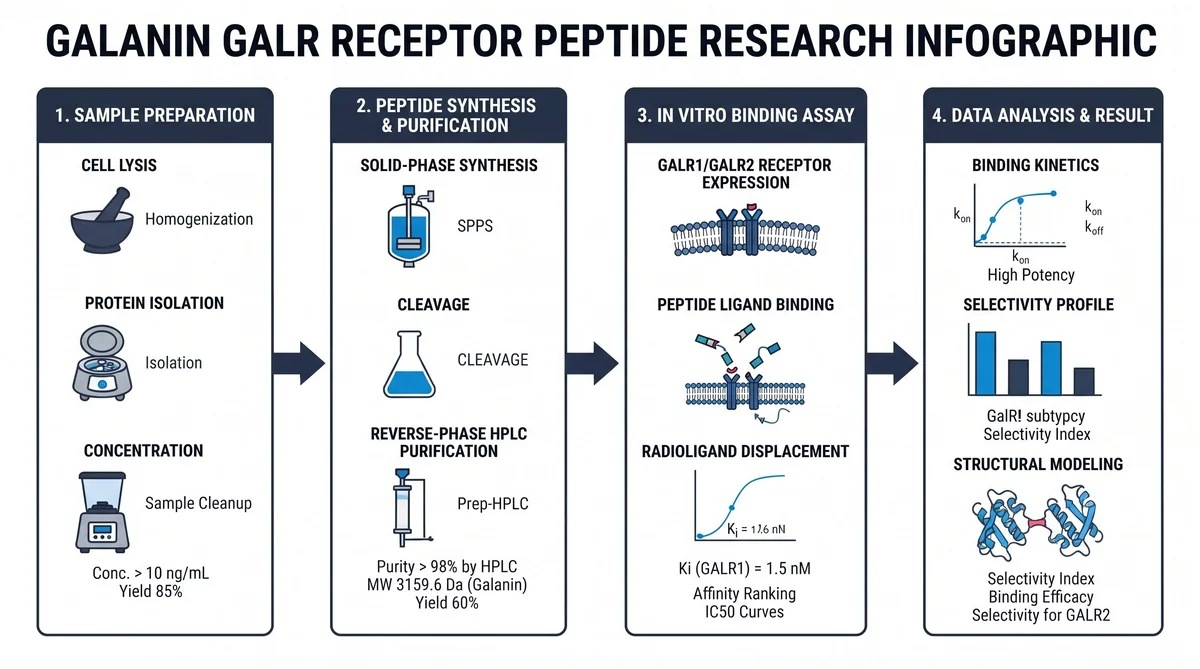
Researchers use labeled analogs to check binding strength at each subtype. A lower Ki value means tighter binding. These measurements help rank compounds by receptor preference.
What signaling pathways do galanin receptors activate?
GalR1 and GalR3 link to Gi/o proteins and block the enzyme that makes cAMP. This lowers cAMP levels inside the target cell. Reduced cAMP slows many downstream cell signals.
GalR2 links to Gq/11 instead and releases calcium from cell stores. It also fires a PKC cascade. This G protein split explains why galanin can drive opposite effects in different tissues.
How is galanin receptor binding affinity measured in vitro?
Binding strength is measured with a labeled-ligand competition assay on cell membrane preps. A labeled galanin analog is added first at a set amount. Then free test peptide is added in rising amounts to compete for the same sites.
The amount that blocks 50 percent of the labeled compound sets the Ki value. A lower Ki means the compound binds more tightly. This gives a standard way to rank test peptides by GalR affinity.
What cell models are used in GalR neuropeptide research?
HEK293 and CHO cell lines are the most common models for GalR receptor work. They can be set up to express just one GalR subtype at a time. This blocks other native receptors and allows clean receptor isolation.
Primary neuron cultures are also used when a closer-to-native setting is needed. These models add biological context that transfected lines lack. Researchers choose based on how much control versus complexity the study requires.
Is galanin involved in sensory signaling research?
Galanin is studied for its role in sensory signal flow at the spinal cord dorsal horn. High peptide levels appear in dorsal root ganglia after nerve stimulation. Studies show it may modulate sensory signals at the spinal level.
According to PubMed (2000), galanin works with other sensory transmitters to shape signal thresholds. These interactions are studied in rodent spinal cord lab models. The data helps map how neuropeptides regulate sensory signal flow.
What is the difference between galanin and GALP?
Galanin-like peptide (GALP) shares part of galanin’s amino acid sequence but has a different tissue spread. While galanin is found widely in the CNS and body tissues, GALP is mainly in the hypothalamus. GALP also has a higher pull toward GalR2 than standard galanin.
Researchers study GALP separately for its role in hypothalamic energy and reproductive signaling. It is not the same as galanin and has its own receptor profile. These differences make it a separate research target.
How is galanin gene expression tracked in lab studies?
Galanin gene expression is tracked with qPCR assays on tissue samples at set time points. Nerve stimulation or hormonal shifts can raise galanin mRNA levels sharply in sensory neurons. Many rodent lab studies have recorded these sharp rises.
By tracking these changes over time, researchers map how the nervous system adjusts neuropeptide output. This shows how the system adapts to stress or external stimuli. The data gives context for interpreting peptide signaling results.
Summary
Galanin is a 29-30 amino acid neuropeptide with three GPCR subtypes: GalR1, GalR2, and GalR3. GalR1 and GalR3 use Gi to lower cAMP. GalR2 uses Gq to raise cell calcium.
Synthetic tools like galnon and M35 let labs isolate each path in cell assays. Future work targets selective ligands for seizure threshold and neurological signaling models.
What Should You Do Next?
Confirm which GalR subtype is active in the signaling path under study. Use HEK293 or CHO cells for clean receptor isolation. Verify that each compound has current HPLC and mass spec data tied to the lot number.
Source peptides at 99% or higher purity to reduce noise in binding assays. For galanin compounds with full lot traceability, visit Next Level Pharm.
People Also Read
- FOXO Pathway Peptide Research: Longevity Signaling
- HIF-1 Alpha Pathway Peptide Research: Hypoxia Studies
About the Author
Next Level Pharm Research Team
The Next Level Pharm research team is composed of biochemists and laboratory scientists dedicated to providing researchers with the highest-purity, lab-tested peptides available. Every batch is HPLC and mass spec verified before dispatch.
Disclaimer: The information provided on this page is for educational and research purposes only. Next Level Pharm’s products are intended for laboratory research use only. They are not intended for human consumption, diagnostic, therapeutic, or medicinal purposes. This content does not constitute medical advice. Always consult a licensed healthcare professional before making any health-related decisions.