
PI3K AKT Pathway Peptide Research: Signaling Studies

Last Updated: October 2026
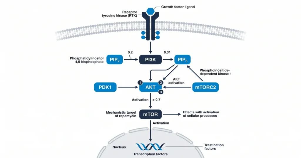
A PI3K AKT pathway peptide research study is an investigation of how molecules activate the phosphoinositide 3-kinase and AKT kinase signaling cascade. PI3K converts PIP2 to PIP3, a second messenger. This step recruits AKT to the membrane. AKT has three isoforms, each with distinct tissue expression. Lab models use synthetic compounds to map how each isoform drives cell survival, growth, and metabolism.
Next Level Pharm provides high-purity research compounds for these lab tasks. The brand keeps 99.4% average purity across its last 100 batches. Each vial is tested by HPLC and mass spec. These protocols give researchers verified materials for their lab work. Every order includes a certificate of analysis. Every batch is third-party tested..
This lab-grade standard lets researchers focus on chemical links. Verified compounds help researchers analyze changes in receptor binding and signal duration. Reliable materials reduce variables in cell culture experiments. This supports cleaner data sets. This approach builds a stable base for studies on cell survival and energy control.
Key Takeaways
- Intracellular Signaling Hub: The PI3K/AKT pathway manages vital cell processes like growth. Survival, and metabolic energy usage. It operates as a complex internal network for cellular information.
- Peptide Trigger Mechanisms: Growth factors like insulin. IGF-1 start the cascade by binding to surface receptors. These ligands activate the receptor tyrosine kinases to begin signaling.
- Central Protein Activation: AKT acts as the main effector protein to trigger key downstream targets like mTORC1. This kinase regulates protein synthesis and cell survival signals.
- Laboratory Quantification Methods: Scientists use Western blotting or ELISA to measure pathway response and p-AKT levels. These tools provide precise data on signaling intensity.
- Aberrant Signaling Relevance: Hyperactive pathways are common in cell proliferation studies. Impaired signaling relates to metabolic issues. Researchers monitor these nodes to understand aberrant signaling states.
These concepts provide a base for exploring the specific mechanisms of cell signals. The next sections examine how pathway components react to external signals and affect cell output.
What is the PI3K/AKT pathway’s role in cell signals?
PI3K/AKT is a central node in cell signals. It transmits cues from surface receptors to cell targets. It links growth factors to a cell’s internal machinery. This cascade controls cell survival, growth, and energy use.
This pathway promotes cell survival by blocking apoptosis. Apoptosis is programmed cell death. Blocking it lets cells keep structural integrity under stress. It also moves glucose transporters to the cell membrane. This boosts energy uptake, because it controls growth and energy use, it is a key research focus.
IGF-1 LR3 is available as a COA-verified research peptide. Every lot is verified by HPLC and mass spec. Researchers monitor how this pathway drives cell cycle progress. This helps them understand basic biology.
How do peptides modulate this specific pathway?
Peptides affect this pathway. They act as upstream activators at RTKs on the cell surface. Growth factors like IGF-1 and insulin are the main ligands. According to the Journal of Peptide Science (2022), this binding event is the first step. Signals move into the cell.
When these peptides bind to RTKs, the receptors self-phosphorylate. This creates docking sites on the inner receptor. The p85 subunit of PI3K binds to these sites. This places the p110 subunit near its substrate at the membrane. This triggers the downstream cascade for cell growth. IGF-1 is available as a COA-verified research peptide. Every lot is verified by HPLC and mass spec.
Which peptides are studied for PI3K/AKT activity?
Peptides studied here include insulin and insulin-like growth factors. Other growth factors also interact with membrane-bound receptors. These proteins start signal cascades by binding to surface receptors. Research focuses on how these ligands shift cell states. Cell models are used.
Synthetic peptide analogs target PI3K/AKT. They selectively activate or block it for lab studies. These molecules allow precise control over receptor binding. IGF-1 LR3 is studied to observe signal effects. It is used without common binding proteins. Researchers use these modified chains to isolate specific nodes.
IGF-1 LR3 is available as a COA-verified research peptide. Every lot is verified by HPLC and mass spec. Altering amino acid sequences shows how signal intensity shifts. Such work clarifies the chemical cues for cell growth.
What are the key downstream effectors of AKT?
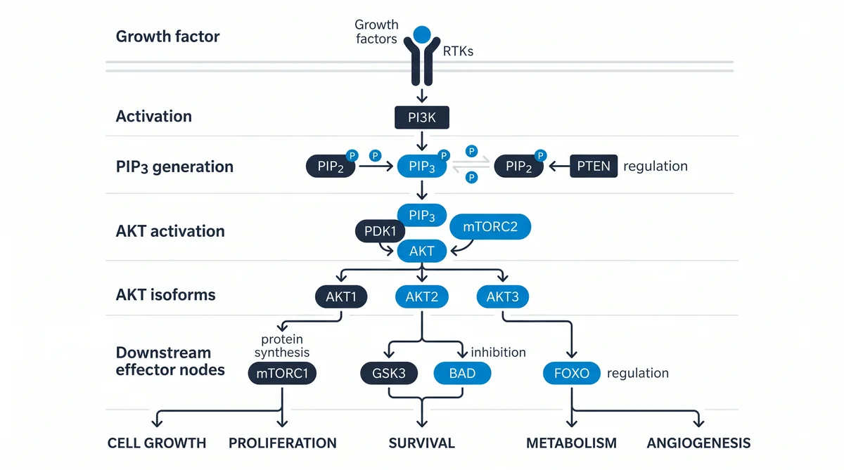
The main AKT effectors include mTORC1, GSK3B, and FOXO factors. AKT phosphorylates these proteins. They direct cell responses to growth signals. They shift metabolism and modify gene expression.
The mTORC1 complex promotes protein synthesis and cell growth. AKT blocks TSC1/TSC2. This lets mTORC1 stimulate ribosome activity and expand cell mass. IGF-1 LR3 is available as a COA-verified research peptide. Every lot is verified by HPLC and mass spec. AKT also blocks GSK3B through phosphorylation. This frees glycogen synthase to support glucose storage. AKT also phosphorylates FOXO factors to push them from the nucleus. This blocks genes that start apoptosis. According to PubMed (2018), this is a key way cells stay alive. High growth signals drive this.
What lab techniques measure pathway response?
Lab techniques track phosphorylation of key proteins. This shows pathway response. They monitor the shift of resting proteins into active states. This shows how ligands affect cell behavior.
Western blotting detects phosphorylated AKT (p-AKT). It separates proteins by size and uses antibodies to find active markers. ELISA kits measure phosphorylated proteins in a sample. For direct enzyme testing, researchers use in vitro kinase assays. These show the rate at which AKT transfers phosphate groups. They show real-time protein activity in the cell. IGF-1 LR3 is available as a COA-verified research peptide. Every lot is verified by HPLC and mass spec.
Why is pathway dysregulation studied in cell models?
PI3K/AKT hyperactivity marks many cell proliferation models. Pathway dysregulation is studied. This abnormal activity promotes uncontrolled growth and blocks apoptosis. Apoptosis is the process of programmed cell death. It is thus a key target for cell growth research. Scientists study this cascade to understand how cells bypass normal growth limits.
Beyond cell proliferation, impaired signals are linked to metabolic signaling disruption. The pathway also affects neuronal signaling models. These involve signal breakdown over time. Researchers study peptides that affect these activities. They look for how balance is restored. IGF-1 LR3 is available as a COA-verified research peptide. Every lot is verified by HPLC and mass spec. According to NCBI (2018), such studies map these shifts. They happen when cell signals fail.
Frequently Asked Questions
What is the PI3K AKT signaling pathway structure?
The PI3K AKT pathway consists of a series of proteins that transmit signals from the cell membrane to the nucleus. It starts with a receptor tyrosine kinase, such as the insulin receptor, which recruits PI3K once it becomes active. PI3K then converts lipids into signaling molecules that recruit AKT to the membrane. This cascade ends by influencing several downstream effector proteins that regulate growth. Protein synthesis, and cellular survival.
How do peptides activate PI3-kinase in lab models?
Peptides activate PI3K by binding to specific surface receptors, which initiates a conformational change in the receptor’s structure. This change triggers autophosphorylation of the intracellular domain. This creates docking sites for the p85 regulatory subunit of PI3K. Once recruited to these sites, the PI3K complex becomes active and begins converting membrane lipids. This process is a common method for observing how extracellular ligands influence internal signaling states in cultured cell models.
What assays measure AKT phosphorylation in vitro?
Researchers frequently use Western blotting to measure the presence of phosphorylated AKT in cell lysates. This method separates proteins by size and uses antibodies to detect specific phosphorylation sites. Alternatively, ELISA or bead-based multiplex assays are used to quantify the concentration of active AKT in a sample. These techniques provide a high level of sensitivity. When assessing the intensity of signaling responses following stimulation with specific peptide or growth factor inputs.
How does the PI3K pathway interact with mTOR signaling?
The PI3K pathway feeds directly into the mTOR signaling network, which acts as a master regulator of cell growth. AKT, once activated, phosphorylates and inhibits the TSC2 protein. This action removes the inhibition on mTORC1. This allows it to become active and start building new proteins. This linkage is a primary research focus for knowledge how cells respond to the availability of nutrients. Growth factors to support structural expansion.
What cell lines are standard for PI3K AKT research?
Researchers often select cell lines that express high levels of growth factor receptors, such. As the MCF-7 breast cell line or the HEK293 kidney cell line. These models are chosen because they possess a well-defined PI3K/AKT signaling architecture. Using shown lines allows for consistent results across different trials. These models are useful for observing how specific experimental conditions shift the balance of cell proliferation. Survival within a controlled lab environment.
Is PI3K upstream or downstream of AKT?
PI3K operates upstream of AKT within this signaling cascade. PI3K drives generating the specific lipid messenger that is required to recruit AKT from the cytoplasm to the plasma membrane. Once AKT is tethered to the membrane by these lipids. Other kinases phosphorylate it to activate its function. Consequently, PI3K activity is a direct requirement for the downstream activation of the AKT protein in most cell types.
What is the function of the PTEN phosphatase?
PTEN acts as a critical negative regulator of the PI3K AKT pathway by converting the signaling lipid PIP3 back into PIP2. This action reduces the amount of PIP3 available at the cell membrane, which prevents the recruitment and activation of AKT. By keeping the pathway in check, PTEN ensures that cell growth. Survival signals do not become hyperactive. Mutations in this gene are often studied to see how pathway dysregulation drives uncontrolled growth.
How does insulin signaling involve the PI3K/AKT pathway?
Insulin signaling relies on this pathway to manage glucose uptake and metabolic energy storage. When insulin binds to its receptor, it stimulates the recruitment of IRS proteins, which then trigger the activation of PI3K and AKT. This sequence of events leads to the translocation of glucose transporters to the cell surface. This allows for the intake of sugar from the surrounding medium. This process is essential for maintaining systemic energy balance in metabolic research models.
Summary
Review your experimental parameters and establish clear control variables for your kinase assays. Confirm that your specific receptor cell lines maintain appropriate expression levels before you initiate the application of your chosen research peptides. High-fidelity data depends on minimizing external interference during the binding phase. So verify your stock concentrations using the provided lot-specific documentation.
Researchers sourcing these signaling-focused peptides can browse the full catalog at Shop. Every vial ships from Next Level Pharm with a COA and full lot traceability.
People Also Read
- BPC-157 Mechanism of Action: A Research-Based Explainer
- What Makes a Peptide Research Grade? Standards and Testing Explained
About the Author
Next Level Pharm Research Team
The Next Level Pharm research team is composed of biochemists and laboratory scientists dedicated to providing researchers with the highest-purity, independently verified peptides available. Every batch is HPLC and mass spec verified before dispatch.
Disclaimer: The information provided on this page is for educational and research purposes only. Next Level Pharm’s products are intended for laboratory research use only. They are not intended for human consumption, diagnostic, therapeutic, or medicinal purposes. This content does not constitute medical advice. Always consult a licensed healthcare professional before making any health-related decisions.