
Transdermal Peptide Delivery Research: Skin Permeability

Last updated: October 2026
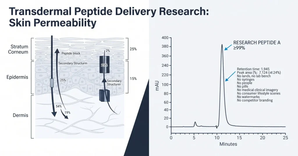
A transdermal peptide delivery study is a lab test that measures how peptides move through skin from the surface to the dermis. The skin barrier blocks most large, charged, and water-loving peptides at the stratum corneum, the outer lipid-rich layer. According to PubMed (2024), peptides above 500 Da cross skin at very low rates without chemical or physical enhancement in lab models.
Next Level Pharm stocks research-grade peptides for lab use, with an average purity of 99.4% across the last 100 batches. Every vial is HPLC and mass spec verified before dispatch. COA records ship with each order for full traceability.
Transdermal delivery research covers three linked areas: skin barrier biology, enhancement methods, and lab tools. Each area informs the others. A study that skips barrier biology will choose the wrong enhancement method.
Key Takeaways
- Skin Barrier: The stratum corneum is a 10-20 micron lipid layer that blocks most peptides by size, charge, and hydrophilicity.
- Molecular Weight Limit: Peptides above 500 Da show poor passive crossing in lab skin models without chemical or physical help.
- Chemical Enhancers: Fatty acids, surfactants, and solvents disrupt lipid packing in the stratum corneum to raise permeation rates.
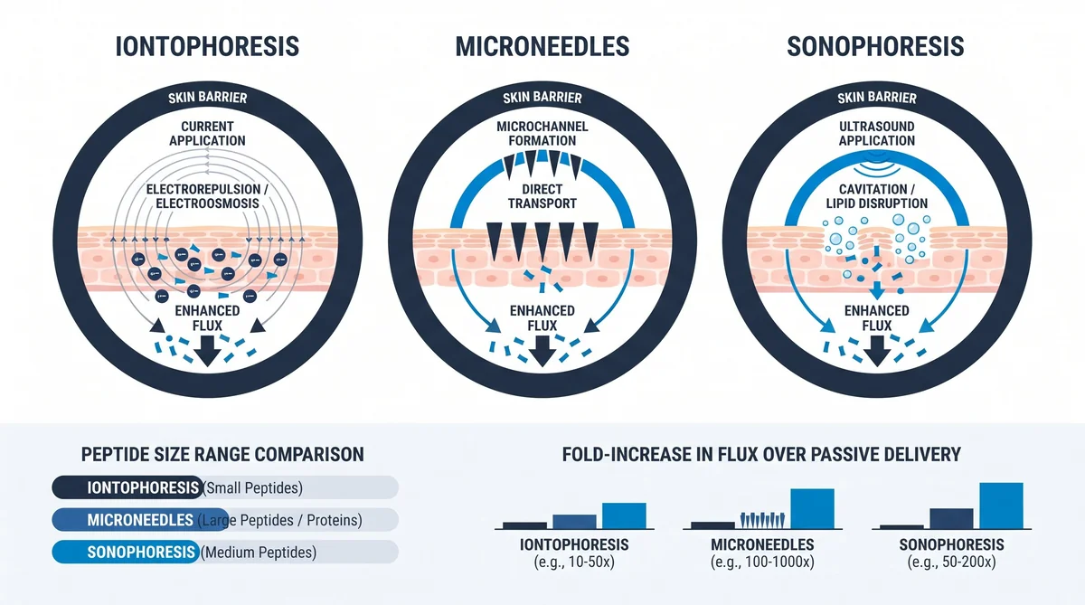
- Physical Methods: Iontophoresis, microneedles, and sonophoresis each drive peptides through the skin layer by a different mechanism.
- Franz Cell Testing: The Franz diffusion cell is the standard lab tool for measuring transdermal flux in excised skin models.
Lab confirmation follows every enhancement test. HPLC quantifies the amount that crossed in each time window. Mass spec confirms the peptide arrived intact on the dermis side.
What Makes the Skin Barrier Hard for Peptides to Cross?
Answer capsule: The stratum corneum is a dense lipid matrix that blocks most peptides. Size, charge, and hydrophilicity each add to the barrier. Very few peptides cross passively without help.
The stratum corneum sits at the top of the epidermis. Its lipid matrix forms a tight seal against water loss. It blocks most external compounds.
Peptides are both large and charged compared to small-molecule drugs. Their MW often exceeds 500 Da. Charged residues also create charge repulsion with the lipid matrix.
Water-loving peptides face a third barrier. The stratum corneum interior is lipid-rich, not aqueous. A hydrophilic compound partitions poorly into this layer and stalls at the surface.
How Does Molecular Weight Affect Transdermal Delivery?
Answer capsule: Higher molecular weight cuts the rate of passive skin crossing. Peptides above 500 Da rarely cross without help. This limit guides which peptides are tested in enhancement studies.
The 500 Da rule holds across most skin models tested in lab settings. Below 500 Da, passive crossing is measurable. Above it, flux drops sharply with each added 100 Da.
According to PubMed (2022), skin crossing for model peptides fell by over 90% as MW rose from 400 to 800 Da in Franz cell studies. The drop was steeper for charged peptides than neutral ones. Neutral analogs showed a smaller but still sharp reduction above 600 Da.
Next Level Pharm supplies COA-verified research peptides across a range of MW values. Labs use these for flux studies. Each batch records purity and MW by mass spec.
What Chemical Methods Enhance Transdermal Permeation?
Answer capsule: Fatty acids, surfactants, and solvents are the main chemical enhancers. Each disrupts the stratum corneum by a different mechanism. Enhancer choice depends on the peptide size and charge.
Fatty acids such as oleic acid fluidize the lipid bilayers inside the stratum corneum. Fluidization opens gaps between corneocytes. Peptides of up to 1 kDa can enter those gaps more readily.
Surfactants disrupt the lipid structure more broadly. Low concentrations raise flux without damaging the membrane. High concentrations cause irreversible damage, so most labs test below 1%.
Solvents such as ethanol act as carriers. They carry peptide into the stratum corneum lipids on contact. The peptide is then in the lipid layer and can diffuse further inward.
| Enhancer Class | Mechanism | Common Example | MW Range |
|---|---|---|---|
| Fatty acids | Lipid fluidization | Oleic acid | Up to 1 kDa |
| Surfactants | Membrane disruption | Polysorbate 80 | Up to 2 kDa |
| Solvents | Carrier penetration | Ethanol | Up to 1.5 kDa |
| Vesicles | Lipid fusion | Liposomes | Up to 5 kDa |
What Physical Methods Aid Transdermal Peptide Study?
Answer capsule: Iontophoresis, microneedles, and sonophoresis are the three main physical methods. Each drives peptides through the skin by a different physical force. Physical methods can reach larger peptides than chemical methods alone.
Iontophoresis applies a low electric current across the skin surface. Charged peptides migrate through the skin with the current. The flux rate depends on charge density and current strength.
Microneedles create tiny channels through the stratum corneum. Peptides enter these channels and diffuse into the dermis below. Channel depth is set by needle length, typically 150 to 900 microns.
According to International Journal of Pharmaceutics (2024), microneedle arrays raised flux for a 2 kDa peptide by 100-fold over passive delivery in porcine skin. Sonophoresis uses low-frequency ultrasound to disrupt the stratum corneum. The cavitation effect opens aqueous channels that close when the field stops.
How Is Transdermal Permeability Measured in Lab Studies?
Answer capsule: The Franz diffusion cell is the standard measurement tool. It holds excised skin between a donor and receiver chamber. HPLC quantifies the peptide that crossed in each time window.
The donor chamber holds the compound on the skin surface. The receiver chamber fills with buffer that mimics tissue fluid. Samples are drawn from the receiver at set time points.
HPLC measures the peak area of the peptide in each receiver sample. The area converts to concentration via a standard curve. A plot of concentration versus time gives the flux curve for the study.
HPLC-verified research peptides help confirm the compound in the donor chamber is pure before the test begins. Impure batches can produce false flux readings. COA records, as described in C-terminal amidation stability research, link purity data to each permeation experiment.
What Peptide Properties Support Transdermal Research?
Answer capsule: Small size, low charge, and moderate lipophilicity help a peptide cross the skin barrier. These three properties define which peptides are good candidates for transdermal studies.
MW below 500 Da is the starting filter for passive studies. Above that, chemical or physical enhancement is always tested first. Very large peptides above 5 kDa are not run in passive Franz cell protocols.
A moderately lipophilic peptide partitions better into the stratum corneum lipid matrix. Extreme lipophilicity causes the peptide to stay in the stratum corneum and not diffuse further. A log P between 0 and 3 covers most well-studied transdermal candidates.
Low total charge helps at neutral skin pH. Cationic peptides can bind to negatively charged skin proteins. That binding reduces flux at low concentrations in standard receiver buffer tests.
Frequently Asked Questions
What makes the skin barrier hard for peptides to cross?
The stratum corneum is a 10-20 micron lipid layer at the skin surface that blocks most peptides by size, charge, and hydrophilicity in lab models. Peptides above 500 Da partition poorly into the lipid matrix and stall at the outer surface before reaching the viable epidermis. Charged residues add a second barrier by interacting with lipid matrix proteins, which slows permeation without chemical or physical enhancement.
How does molecular weight affect transdermal peptide delivery?
Peptides below 500 Da show measurable passive flux in Franz cell studies, while those above show flux that drops sharply with each added 100 Da. According to PubMed (2022), model peptide permeation fell by over 90% as MW rose from 400 to 800 Da in controlled Franz cell experiments. The 500 Da guideline is the first filter applied when selecting peptides for transdermal delivery studies in a research context.
What chemical methods are used to enhance transdermal permeation?
Fatty acids such as oleic acid fluidize the stratum corneum lipids to open gaps for peptide entry at low concentrations. Surfactants disrupt the lipid layer more broadly, with concentrations below 1% raising flux while higher levels cause irreversible barrier damage. Solvents such as ethanol act as carriers, partitioning peptides into the stratum corneum lipids on contact and enabling further inward diffusion.
What physical methods are used in transdermal peptide research?
Iontophoresis drives charged peptides through skin by applying a low electric current, where flux rate depends on charge density and current strength. Microneedles create channels through the stratum corneum that allow peptides up to 5 kDa to reach the dermis by direct diffusion. Sonophoresis uses ultrasound cavitation to open temporary aqueous channels in the stratum corneum, which close after the ultrasound field stops.
How is transdermal permeability measured in lab studies?
The Franz diffusion cell holds excised skin between a donor and receiver chamber, with samples drawn at set time points to build a flux curve. HPLC quantifies the peptide in each receiver sample by converting peak area to concentration through a calibration standard curve. Mass spec confirms the peptide arrived intact without degradation, since a fragment peak in the receiver would indicate enzymatic breakdown during the crossing.
What peptide properties support transdermal delivery research?
MW below 500 Da, a log P of 0-3, and low charge at neutral pH are the three filters for passive transdermal research candidates. A peptide that meets all three can cross the stratum corneum passively at measurable rates in standard excised skin models. Peptides outside this window still serve as research subjects but require iontophoresis, microneedles, or chemical enhancement to generate detectable flux.
Why does the 500 Da rule matter in transdermal research?
The 500 Da threshold is the size above which passive crossing through the stratum corneum becomes too low to measure in standard Franz cell setups. Peptides near this size are the first target for enhancement studies, as a small flux rise from a low baseline is easier to detect. Those well above it (above 2-3 kDa) require physical methods rather than chemical ones to generate any meaningful permeation signal.
How is peptide integrity confirmed after transdermal delivery studies?
Mass spec on the receiver samples confirms the compound that crossed is the intact peptide, not a fragment from protease activity. HPLC confirms concentration but cannot distinguish intact peptide from closely eluting fragments, so mass spec is the required second check. Next Level Pharm provides COA records with mass spec data for every batch, so labs can document starting purity and compare it to receiver results.
Summary
Transdermal peptide delivery research maps how compounds move through the stratum corneum into deeper skin layers. The barrier blocks most peptides by size, charge, and hydrophilicity. Understanding these three properties is the first step before choosing an enhancement method.
Chemical enhancers such as fatty acids, surfactants, and solvents disrupt the lipid matrix to raise flux rates. Physical tools including iontophoresis, microneedles, and sonophoresis drive compounds through the barrier by direct force. Franz cell studies with HPLC and mass spec confirm how much peptide crossed and in what form.
Researchers choose compounds based on MW, charge, and log P before any lab run. Properties outside the passive window call for enhancement from the start. COA-confirmed purity at the source reduces variables in any permeation study.
What Should You Do Next?
Select peptides based on MW, charge, and log P before designing a transdermal study protocol. Run Franz cell tests with both passive and enhanced conditions to bracket the permeation window. Next Level Pharm provides COA-confirmed research peptides and HPLC purity records for all transdermal flux research needs.
People Also Read
- Sublingual Peptide Absorption: What Studies Show
- Why Most Peptides Fail Orally: Degradation Research
About the Author
Next Level Pharm Research Team
The Next Level Pharm research team is made up of biochemists and lab scientists. Their goal is to give researchers the highest-purity, COA-confirmed compounds available. Every batch is HPLC and mass spectrometry tested before dispatch.
Disclaimer: The information provided on this page is for educational and research purposes only. Next Level Pharm’s products are intended for laboratory research use only. They are not intended for human consumption, diagnostic, therapeutic, or medicinal purposes. This content does not constitute medical advice. Always consult a licensed healthcare professional before making any health-related decisions.