
HIF-1 Alpha Pathway Peptide Research: Hypoxia Studies

Last Updated: October 2026
HIF-1 alpha pathway peptide research maps how small proteins interact with cellular oxygen sensors. These sensors regulate which genes activate under low-oxygen conditions. According to PubMed (2024), eight HIF-1 alpha target genes serve as activity markers in cultured cells. Researchers use these genes to track signal state across multiple cell types.
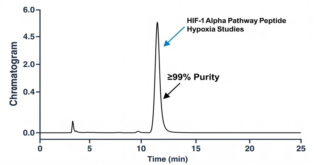
Next Level Pharm offers COA-verified research peptides for lab investigations. Each vial carries a lot-specific certificate of analysis. HPLC and mass spectrometry confirm purity at 99.4% average across the last 100 batches. The catalog includes compounds relevant to oxygen-sensing and mitochondrial research.
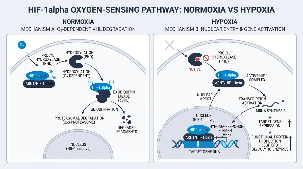
The HIF-1 alpha hypoxia pathway involves a regulated series of protein interactions. Under normal oxygen, PHD enzymes add hydroxyl groups to proline residues on HIF-1 alpha. This step allows VHL (von Hippel-Lindau tumor suppressor protein) to bind HIF-1 alpha. VHL then triggers breakdown via the proteasome, the cell’s protein-disposal system. When oxygen falls, PHD activity drops and HIF-1 alpha accumulates in the cell nucleus.
Key Takeaways
1. Core Oxygen Sensor: HIF-1 alpha is the primary transcription factor for cellular hypoxia responses. It coordinates gene programs for oxygen delivery and metabolic adaptation.
2. PHD as the Switch: PHD (prolyl hydroxylase domain) enzymes act as the pathway’s oxygen-dependent switch. They require molecular oxygen to tag HIF-1 alpha for destruction.
3. VHL as the Gatekeeper: VHL protein binds hydroxylated HIF-1 alpha and recruits an E3 ubiquitin ligase complex. This marks HIF-1 alpha for proteasomal degradation.
4. Peptide Interaction Points: Peptides studied in vitro can bind the ODD (oxygen-dependent degradation) domain of HIF-1 alpha. Some block PHD-mediated hydroxylation. Others block VHL recognition.
5. Assay Readouts: Luciferase reporter assays and Western blotting measure pathway activity in lab models. They track protein levels and transcriptional output.
6. Cell Model Choices: HCT116 colon cancer cells and HEK293 kidney cells are common research platforms. Both carry well-characterized hypoxia signaling architecture.
These mechanisms form the foundation of HIF-1 alpha pathway peptide research. The next sections examine each component for laboratory researchers.
What Is the HIF-1 Alpha Hypoxia Response Pathway?
HIF-1 alpha (hypoxia-inducible factor 1 alpha) is part of a two-subunit transcriptional complex. It pairs with HIF-1 beta to bind HRE (hypoxia response element, a DNA sequence near oxygen-regulated genes) in target gene promoters. Under normal oxygen, PHD enzymes prevent this complex from forming. Under low oxygen, the complex activates genes for glucose transport and metabolic adaptation.
Three regulatory nodes control the pathway. First, PHD-mediated hydroxylation tags HIF-1 alpha for proteasomal clearance. Second, VHL protein recognizes the tagged form and recruits a ligase complex. Third, FIH-1 (factor inhibiting HIF-1, an asparagine hydroxylase enzyme) blocks HIF-1 alpha transactivation under normoxia. Each node is a distinct research target for peptide interaction studies.
- PHD Hydroxylation: PHD2 adds a hydroxyl group to Pro402 and Pro564 within the ODD domain.
- VHL Recognition: VHL reads the hydroxylated prolines and links to the ubiquitin machinery.
- FIH-1 Repression: FIH-1 modifies Asn803 to block coactivator binding at the transactivation domain.
How Do Peptides Stabilize or Destabilize HIF-1 Alpha?
Peptides interact with the HIF-1 alpha pathway at multiple points. Cyclic and linear peptides can block the PHD-substrate interaction in cell-free assays. Others compete for VHL binding. A third class targets the HIF-1 alpha and HIF-1 beta dimerization interface. According to PubMed (2013), a cyclic peptide targeting HIF-1 heterodimerization showed measurable inhibition of hypoxia signaling in cell culture models.
Stabilizing peptides use sequences from the ODD domain. These compete with the native protein for PHD2 active site access. They serve as probes for mapping enzyme-substrate contacts. Destabilizing peptides enhance PHD-VHL assembly by acting as scaffold molecules. According to PMC (2023), protein scaffolds influence HIF-1 alpha turnover rates under chronic hypoxia.
What Assays Measure HIF-1 Alpha Activity in Vitro?
Lab methods quantify HIF-1 alpha activity at different pathway layers. Multiple complementary approaches are needed for full characterization. According to PubMed (2024), combining two or more assay types gives the most robust activity profile.
- Western Blotting: Detects HIF-1 alpha protein in cell lysates using specific antibodies. It gives a direct measure of protein abundance.
- Luciferase Reporter Assay: An HRE-driven luciferase construct measures transcriptional activity. Luminescence output reflects active HIF complex binding at gene promoters.
- ELISA: Enzyme-linked immunosorbent assay kits quantify HIF-1 alpha protein concentration from cell extracts. These kits return numerical readouts for comparing treatment conditions.
- qPCR for Target Genes: Quantitative PCR measures mRNA levels of known pathway targets. VEGF (vascular endothelial growth factor) and GLUT1 (glucose transporter 1, a cell-surface sugar transporter) are common endpoints.
- PHD Activity Assay: A cell-free test monitors product formation as PHD enzymes process a short ODD-domain peptide. This directly measures enzyme kinetics.
A research note: SS-31 is a mitochondria-targeting research peptide available with COA and lot documentation. Its research intersects with oxygen-sensing biology through mitochondrial membrane potential studies.
How Does PHD Enzyme Regulate HIF-1 Alpha Degradation?
PHD enzymes (prolyl hydroxylase domain enzymes, a family of dioxygenases that use oxygen as a co-substrate) are the primary oxygen sensors in this pathway. Three isoforms exist: PHD1, PHD2, and PHD3. PHD2 is the main regulator in most cell types. These enzymes require molecular oxygen, iron(II), and 2-oxoglutarate to function. Without any co-factor, hydroxylation stalls, so iron chelation and hypoxia both stabilize HIF-1 alpha in lab models.
PHD2 hydroxylates Pro564 in the ODD domain. This step is rate-limiting for the entire degradation sequence. VHL then binds the hydroxylated proline through a defined groove on its beta-domain. This recruits Elongin B, Elongin C, and a Cullin-2-based E3 ligase. The ligase attaches ubiquitin chains to HIF-1 alpha for proteasomal destruction. According to PMC (2017), PHD-dependent degradation accounts for the majority of HIF-1 alpha turnover under normoxic conditions. Short ODD-domain peptides map the exact VHL binding residues and serve as positive controls in competitive binding assays.
What Cell Models Are Used for Hypoxia Research?
Cell model selection shapes assay reproducibility and data interpretation. Researchers choose lines based on known HIF-1 alpha signaling architecture. Consistent expression levels are key for cross-experiment comparisons.
- HCT116 Cells: A human colon carcinoma line with high basal HIF-1 alpha expression and a well-defined signaling context.
- HEK293 Cells: A human embryonic kidney line. Easy transfection and low background HIF-1 alpha support stable reporter studies.
- PC12 Cells: A rat adrenal pheochromocytoma line used in PHD inhibitor studies for neural-context oxygen-sensing responses.
- HUVEC Cells: Human umbilical vein endothelial cells, selected when angiogenic outputs like VEGF are the primary endpoint.
- MCF-7 Cells: A human breast cancer line used in HIF-1 alpha overexpression studies within cancer biology contexts.
BPC-157 and SS-31 are both available at the Next Level Pharm recovery catalog. Both are studied in cellular stress and mitochondrial biology models, areas that overlap with oxygen-sensing research.
How Does VHL Protein Interact with HIF-1 Alpha?
VHL (von Hippel-Lindau protein) is the substrate recognition subunit of an E3 ubiquitin ligase complex. It binds HIF-1 alpha only after PHD-mediated hydroxylation at Pro564 and Pro402. A specific binding groove on the VHL beta-domain accommodates the hydroxyproline residue with high selectivity. Loss-of-function mutations in VHL result in constitutive HIF-1 alpha accumulation. This is a key mechanistic finding in renal cell carcinoma research models.
Short hydroxyproline-containing peptides from the ODD region have been crystallized with VHL, defining the contact surface at the atomic level. A 2023 study in PMC showed that SIRT1-mediated signaling can alter VHL recruitment efficiency. This adds a metabolic layer to classical oxygen-sensing models. Such data expand the toolkit for researchers studying oxygen and energy status together in vitro.
| Component | Oxygen Condition | Activity | Effect on HIF-1 Alpha |
|---|---|---|---|
| PHD2 enzyme | Normoxia (21% O2) | High | Hydroxylates Pro564; triggers VHL binding |
| PHD2 enzyme | Hypoxia (less than 1% O2) | Low or absent | No hydroxylation; HIF-1 alpha accumulates |
| VHL protein | Normoxia | Active | Recruits E3 ligase; HIF-1 alpha degraded |
| VHL protein | Hypoxia | Inactive | HIF-1 alpha escapes degradation |
| FIH-1 enzyme | Normoxia | Active | Blocks coactivator binding; limits gene output |
| FIH-1 enzyme | Hypoxia | Inactive | HIF-1 alpha and HIF-1 beta complex activates genes |
| HIF-1 alpha | Normoxia | Rapidly degraded | Half-life less than 5 minutes |
| HIF-1 alpha | Hypoxia | Stabilized | Translocates to nucleus; activates target genes |
Frequently Asked Questions
What Is the HIF-1 Alpha Hypoxia Response Pathway?
The HIF-1 alpha (hypoxia-inducible factor 1 alpha) pathway is a cellular oxygen-sensing system. Under normal oxygen, PHD enzymes hydroxylate two proline residues in HIF-1 alpha. This modification triggers VHL binding and ubiquitin-mediated destruction via the proteasome. When oxygen falls, PHD activity declines and HIF-1 alpha escapes destruction. The stabilized protein enters the nucleus, pairs with HIF-1 beta, and activates genes that govern oxygen delivery, glucose uptake, and cell survival under low-oxygen conditions.
How Do Peptides Stabilize or Destabilize HIF-1 Alpha?
Peptides modulate HIF-1 alpha stability by targeting specific protein-protein or enzyme-substrate contacts. Some compete with the native HIF-1 alpha ODD domain at the PHD2 active site, which lowers hydroxylation efficiency. Others block the VHL binding groove to prevent recognition of the hydroxylated form. A third group targets the dimerization interface. Published research documents both stabilizing and destabilizing peptide classes as useful probes for mapping interaction surfaces in cell-free and cell-based experiments.
What Assays Measure HIF-1 Alpha Activity in Vitro?
Western blotting and luciferase reporter assays are the most common approaches. Western blotting detects HIF-1 alpha protein in cell lysates using subunit-specific antibodies. Luciferase reporters use an HRE promoter linked to a luminescent enzyme. ELISA kits provide a quantitative protein concentration readout. PHD activity can also be measured in cell-free systems using a short ODD-peptide substrate. Combining two or more of these methods gives the most robust characterization of pathway activity.
How Does PHD Enzyme Regulate HIF-1 Alpha Degradation?
PHD (prolyl hydroxylase domain) enzymes are dioxygenases that use molecular oxygen as a co-substrate. They hydroxylate two proline residues within the ODD domain of HIF-1 alpha, creating a high-affinity site for VHL binding. PHD2 is the primary isoform in most tissues. It requires iron(II), oxygen, and 2-oxoglutarate to catalyze the reaction. Under hypoxia or iron depletion, the reaction stalls. HIF-1 alpha then accumulates because it cannot be delivered to the VHL and ubiquitin machinery.
What Cell Models Are Standard for Hypoxia Signaling Research?
HCT116 colon carcinoma cells and HEK293 kidney cells are the most widely used platforms. Both carry well-characterized HIF-1 alpha signaling architecture. HCT116 cells have been used to create endogenous NanoLuc reporter alleles for real-time tracking. HEK293 cells accept stable HRE-luciferase constructs with high efficiency. HUVEC (human umbilical vein endothelial cells) are selected when vascular gene outputs like VEGF are the study endpoint. PC12 cells offer a neural-context model for PHD inhibitor studies.
What Role Does VHL Protein Play in HIF-1 Alpha Stability?
VHL (von Hippel-Lindau protein) is the recognition subunit of an E3 ubiquitin ligase complex. It binds HIF-1 alpha only after PHD enzymes add a hydroxyl group to specific proline residues. Once VHL binds, it recruits Elongin B and Elongin C, which brings a Cullin-2-based ligase to attach ubiquitin chains. The tagged protein is then delivered to the proteasome. Mutations in VHL disrupt this process and result in constitutive HIF-1 alpha accumulation in research models.
What Downstream Genes Does HIF-1 Alpha Activate?
HIF-1 alpha activates genes that support cell survival under low-oxygen conditions. Key targets include VEGF (vascular endothelial growth factor, a protein that stimulates blood vessel formation) and GLUT1 (glucose transporter 1). Other targets include LDHA (lactate dehydrogenase A, an enzyme that converts pyruvate to lactate) and EPO (erythropoietin, a protein that signals red blood cell production). Each target carries an HRE sequence in its promoter region. HIF-1 alpha binds these sequences together with HIF-1 beta to activate transcription.
How Does Oxygen Concentration Affect HIF-1 Alpha Half-Life?
Under normoxia (21% oxygen), HIF-1 alpha has a half-life of less than 5 minutes. PHD2 activity is high and the degradation cycle runs continuously. As oxygen falls below 5%, PHD2 activity declines in proportion. Below 1% oxygen, the half-life extends to over 30 minutes. Cobalt chloride and iron chelators mimic hypoxia by inhibiting PHD enzymes in normoxic conditions. These chemical tools serve as controls in assay development studies.
What Is the FIH-1 Enzyme and How Does It Function?
FIH-1 (factor inhibiting HIF-1, an asparagine hydroxylase enzyme) modifies Asn803 in the C-terminal transactivation domain of HIF-1 alpha. This modification prevents p300 and CBP coactivator proteins from binding. These coactivators are required for full transcriptional output at HRE-containing genes. FIH-1 uses molecular oxygen but has a lower affinity for oxygen than PHD2. The result is a graded response: moderate hypoxia allows HIF-1 alpha to accumulate but still limits its transcriptional output.
Which Peptides Have Been Studied as HIF-1 Alpha Research Probes?
Several peptide classes appear in the HIF-1 alpha research literature. ODD-domain peptides containing hydroxyproline serve as high-affinity VHL ligands and competitive binding tools. Cyclic peptides targeting the HIF-1 alpha and HIF-1 beta dimerization interface have been tested in cell culture models. Multivalent peptides on scaffold backbones have been studied for enhanced potency at the VHL binding groove. These probes help map interaction surfaces and study enzyme-substrate kinetics. All documented studies are conducted in biochemical assay formats.
Summary
HIF-1 alpha (hypoxia-inducible factor 1 alpha) is the primary regulator of cellular oxygen-response gene programs. The pathway is controlled by PHD enzymes (prolyl hydroxylase domain enzymes), VHL protein, and FIH-1. These regulators keep HIF-1 alpha near zero under normal oxygen. When oxygen falls, the system shifts and HIF-1 alpha drives metabolic adaptation genes. Peptide probes from the ODD domain and dimerization interface map enzyme-substrate contacts in lab models. Assay methods range from luciferase reporters to PHD activity kits, with HCT116 and HEK293 providing reproducible in vitro platforms for hypoxia research.
What Should You Do Next?
Lab researchers studying the HIF-1 alpha pathway need compounds with documented purity before starting assays. Contaminants can produce false signals at PHD or VHL binding sites. Lot traceability also supports reproducibility checks across experiments.
Shop research peptides at Next Level Pharm for HPLC-tested, mass spec-verified compounds. Every batch is third-party tested.. Explore the recovery peptide catalog for compounds relevant to cellular stress and mitochondrial research models.
People Also Read
- SS-31 Research: Mitochondria-Targeting Peptide
- BPC-157 Mechanism of Action: A Research-Based Explainer
About the Author
Next Level Pharm Research Team
The Next Level Pharm research team is composed of biochemists and laboratory scientists dedicated to providing researchers with the highest-purity, independently verified peptides available. Every batch is HPLC and mass spec verified before dispatch.
Disclaimer: The information provided on this page is for educational and research purposes only. Next Level Pharm’s products are intended for laboratory research use only. They are not intended for human consumption, diagnostic, therapeutic, or medicinal purposes. This content does not constitute medical advice. Always consult a licensed healthcare professional before making any health-related decisions.