
PNC-27 Research: p53 MDM2-Binding Domain Peptide Studies

Last updated: June 2026
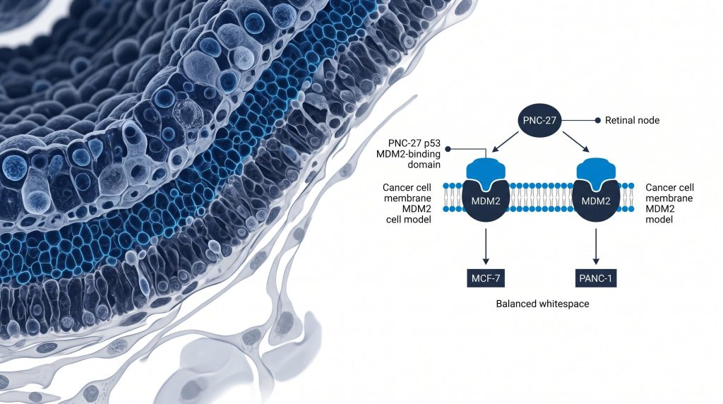
A PNC-27 research compound is a synthetic peptide derived from residues 12-26 of the tumor suppressor protein p53. It is fused to a penetratin transmembrane sequence that directs it toward cell membranes. Researchers study PNC-27 in cancer cell line models to examine how the peptide interacts with MDM2 expressed on the plasma membrane. Cell lines used in published studies include MCF-7, PANC-1, and MDA-MB-231.
Next Level Pharm is a US-based supplier of research-grade PNC-27, verified to ≥99.4% purity by HPLC. Mass spectrometry on every batch. Each lot ships with a COA. A lot number for full traceability.
Key Takeaways
- PNC-27 contains the p53 MDM2-binding domain. PNC-27 is p53 residues 12-26 fused to a penetratin transmembrane sequence. MDM2 is overexpressed on the plasma membrane of certain cancer cell lines.
- MDM2 expression differs by cell type. MDM2 is present on the plasma membrane of MCF-7, PANC-1. MDA-MB-231 cancer cells. It is absent from the plasma membrane of normal, non-transformed cells.
- LDH and PI assays measure membrane disruption. Lactate dehydrogenase (LDH) release. Propidium iodide (PI) staining are the primary readouts for PNC-27 membrane activity in cell studies.
- COA-verified lots: Next Level Pharm ships lyophilized PNC-27 lots with HPLC. Mass spec data on every batch. Each lot has a traceable number.
- Research use only. All PNC-27 in this catalog is for lab research use only. No clinical outcomes are claimed.
What Is PNC-27 and How Is It Structured?
PNC-27 is a synthetic peptide containing p53 residues 12-26 fused to a penetratin transmembrane sequence. According to Bowne et al. (2008), PNC-27 disrupts the plasma membranes of cancer cells that overexpress MDM2. MDM2 (murine double minute 2) is an E3 ubiquitin ligase. In normal cells, MDM2 is found primarily in the nucleus and cytoplasm. In certain cancer cell lines, MDM2 is also present on the outer plasma membrane.
The p53 sequence in PNC-27 covers residues 12-26. This region is the natural MDM2-binding domain of p53. The penetratin sequence is a cell-penetrating peptide (CPP) derived from the Drosophila Antennapedia homeodomain. It enables PNC-27 to associate with lipid bilayers.
When PNC-27 encounters cells that display MDM2 on their plasma membrane, it binds MDM2 at the cell surface. This binding event is studied using MDM2 cell staining and co-location assays. Normal human fibroblasts. Epithelial cells are used as negative controls because they lack plasma membrane MDM2.
| Cell Line | MDM2 Membrane Expression | PNC-27 Binding | LDH Release |
| MCF-7 | High | Yes | Elevated |
| PANC-1 | High | Yes | Elevated |
| MDA-MB-231 | High | Yes | Elevated |
| Normal fibroblasts | Absent | No | Baseline |
What Role Does MDM2 Play in PNC-27 Cell Studies?
MDM2 overexpression on the plasma membrane of cancer cells is the key requirement for PNC-27 activity in cell models. According to Patel et al. (2011), MDM2 overexpression is a common feature of multiple cancer cell lines. MDM2 normally acts as a negative regulator of p53. It binds p53. Marks it for proteasomal degradation. Many cancer lines amplify the MDM2 gene to suppress p53-mediated cell cycle arrest.
MDM2 membrane location is distinct from its nuclear function. Several research groups have identified MDM2 on the plasma membrane of cancer cells using cell staining and flow flow assay. The mechanism of membrane targeting is not fully characterized but may involve post-translational modifications.
In PNC-27 studies, MDM2 cell staining is used to confirm membrane expression before applying the peptide. This step is critical for interpreting LDH release data. If a cell line does not express plasma membrane MDM2, PNC-27 shows minimal LDH signal above background levels.
Researchers use nutlin-3 (a small-molecule MDM2 inhibitor) as a mechanistic comparator. Nutlin-3 blocks the p53 MDM2 interaction in the nucleus, not at the plasma membrane. This distinction separates the mechanism of action from classical MDM2 inhibitors.
Which Cell Lines Are Used in PNC-27 Research?
MCF-7, PANC-1. MDA-MB-231 are the primary cell lines used in PNC-27 research. According to Hilchie et al. (2013), anticancer peptides that act at the plasma membrane require cell lines with confirmed membrane target expression for valid assay results. MCF-7 is a human breast adenocarcinoma line. It has high MDM2 expression. Is widely used in membrane-active peptide studies.
PANC-1 is a human pancreatic carcinoma line with high MDM2 expression. MDA-MB-231 is a triple-negative breast cancer line. It is more invasive than MCF-7. Is used to study PNC-27 activity in a more aggressive cancer cell model.
Normal cell controls are required in every PNC-27 experiment. Primary human dermal fibroblasts. Or hUVEC endothelial cells are common negative controls. These cell types lack plasma membrane MDM2. Serve as target range benchmarks.
All PNC-27 studies described here are in vitro cell model research only. No clinical outcomes are implied or claimed. This research does not establish safety. Or benefit in humans.
How Is PNC-27 Activity Measured in Cell Assays?
LDH release, PI staining, MDM2 cell staining. Caspase-3/7 activity are the four primary PNC-27 endpoints. LDH (lactate dehydrogenase) release is the main cell death endpoint in PNC-27 research. LDH is a cytoplasmic enzyme. Its presence in the culture medium indicates membrane rupture. LDH release assays use colorimetric kits with absorbance at 490 nm. Results are expressed as percent cell death relative to total LDH control.
PI (propidium iodide) staining is a complementary membrane integrity assay. PI is a cell-impermeant dye. It enters cells only when the plasma membrane is compromised. Flow flow assay or fluorescence microscopy quantifies PI-positive cells. High PI uptake confirms membrane disruption.
MDM2 cell staining uses a primary anti-MDM2 antibody. A fluorescent secondary antibody. Confocal microscopy identifies co-location of MDM2 signal with the plasma membrane marker. This confirms MDM2 surface expression before PNC-27 treatment.
Caspase-3/7 activity is measured with the Caspase-Glo 3/7 assay to track whether apoptosis follows membrane disruption. Researchers run LDH. Caspase-3/7 on the same time-course to understand the sequence of cellular events.
How Does PNC-27 Compare to Other MDM2 Research Tools?
PNC-27 acts at the plasma membrane, while nutlin-3. P53 peptide 12-26 act inside cells. Three compounds are commonly compared in MDM2 research. PNC-27, the p53 peptide fragment (residues 12-26 alone), and nutlin-3. The table below summarizes their key differences:
| Compound | Structure | Site of Action | MDM2 Interaction | Key Assay |
| PNC-27 | p53(12-26) + penetratin | Plasma membrane | MDM2 on membrane | LDH release |
| p53 peptide (12-26) | p53 sequence alone | Intracellular | Nuclear MDM2 | Co-IP |
| Nutlin-3 | Small molecule | Intracellular | MDM2-p53 binding groove | p53 protein level |
The penetratin sequence is the key structural element that directs PNC-27 to the plasma membrane. Without it, the p53 12-26 fragment cannot access surface MDM2. This makes PNC-27 mechanistically distinct from both nutlin-3. The unmodified p53 peptide.
Browse recovery research peptides for PNC-27 lots with COA. Mass spec data.
What Research Tools Are Available for PNC-27 Studies?
COA-verified PNC-27 lots alongside LDH assay kits. MDM2 antibodies form the core toolkit for membrane peptide cell studies.
Research teams need a verified PNC-27 lot to run any study. The lot must match the published reference standard for molecular weight and purity. Next Level Pharm verifies each PNC-27 lot by HPLC. Mass spectrometry before shipping. Common assay kits include the CytoTox 96 LDH kit, PI flow flow assay reagents. The Caspase-Glo 3/7 assay for downstream apoptosis measurement. A validated anti-MDM2 antibody (clone SMP14. Or 2A10) is required for plasma membrane MDM2 cell staining confirmation. View the PNC-27 product page for lot-specific COA. Mass spec data.
Frequently Asked Questions
What is PNC-27 in peptide research?
PNC-27 is a synthetic peptide containing p53 residues 12-26 fused to a penetratin transmembrane sequence. Researchers use it to study how the p53 MDM2-binding domain interacts with MDM2 expressed on the plasma membrane of cancer cell lines. Published in vitro studies have used MCF-7, PANC-1. MDA-MB-231 cancer cell models. PNC-27 is a research compound for lab research use only.
What is MDM2 and why is it studied in cancer cell lines?
MDM2 (murine double minute 2) is an E3 ubiquitin ligase that normally binds and degrades p53. In many cancer cell lines, the MDM2 gene is amplified, suppressing p53-mediated cell cycle arrest. A subset of cancer cell lines also expresses MDM2 on the outer plasma membrane, not just in the nucleus or cytoplasm. This membrane location is the specific target studied in PNC-27 research.
What does the penetratin sequence do in PNC-27?
Penetratin is a cell-penetrating peptide (CPP) derived from the Drosophila Antennapedia homeodomain. In PNC-27, the penetratin sequence is fused to the p53 12-26 MDM2-binding domain. This fusion directs the peptide toward lipid bilayers. Enables it to access MDM2 on the outer plasma membrane of cancer cells. Without the penetratin segment, the p53 fragment alone cannot reach surface MDM2.
What is the LDH release assay in PNC-27 studies?
The LDH (lactate dehydrogenase) release assay measures cell death by detecting the cytoplasmic enzyme LDH in culture medium. LDH is released when the plasma membrane is disrupted. The assay uses a colorimetric kit read at 490 nm. Results are expressed as percent cell death. In PNC-27 cancer cell studies, elevated LDH signal indicates membrane disruption in MDM2-positive cell lines.
What is propidium iodide staining used for in PNC-27 research?
Propidium iodide (PI) is a cell-impermeant fluorescent dye. It enters cells only when the plasma membrane is compromised. Flow flow assay or fluorescence microscopy counts PI-positive cells. In PNC-27 studies, PI staining is run alongside LDH release to confirm membrane disruption at the single-cell level. Together, LDH and PI provide complementary evidence for PNC-27 membrane activity in cancer cell lines.
How does PNC-27 differ from nutlin-3?
PNC-27 is a peptide that acts at the plasma membrane by binding MDM2 expressed on the cancer cell surface. Nutlin-3 is a small molecule that works inside cells by blocking the MDM2-p53 binding groove in the nucleus. These are distinct mechanisms of action. PNC-27 and nutlin-3 are used in parallel as research comparators to distinguish membrane-level from intracellular MDM2 interactions in cell studies.
What normal cell controls are used in PNC-27 experiments?
Primary human dermal fibroblasts. HUVEC endothelial cells are common normal cell controls in PNC-27 research. These cell types lack plasma membrane MDM2 expression. They serve as target range benchmarks. A well-designed PNC-27 experiment shows elevated LDH in MDM2-positive cancer lines but baseline LDH in normal cell controls. Including these controls is essential for interpreting PNC-27 activity data correctly.
Is PNC-27 approved for clinical or therapeutic use?
No. PNC-27 is a research-use-only peptide compound. All published data is from in vitro cell culture models. PNC-27 is not approved by the FDA for any clinical or therapeutic application. It is not intended for clinical use. Researchers should review all applicable institutional biosafety. Ethical guidelines before conducting any study with PNC-27. Or related research compounds.
Summary
PNC-27 research focuses on a synthetic peptide containing p53 residues 12-26 fused to a penetratin transmembrane sequence. It is studied in MCF-7, PANC-1. MDA-MB-231 cancer cell lines that overexpress MDM2 on the plasma membrane. Normal cells lacking plasma membrane MDM2 serve as negative controls in every experiment.
Key endpoints in PNC-27 cell studies include LDH release, PI staining, MDM2 cell staining, and caspase-3/7 activity. These assays provide complementary evidence for membrane disruption and downstream events. Nutlin-3 and the unmodified p53 12-26 fragment are used as mechanistic comparators in well-designed studies.
PNC-27 is available as a COA-verified, lyophilized research lot. HPLC and mass spec data confirm purity on every batch. This is a research-grade compound only. Is not intended for clinical use.
What Should You Do Next?
- Review the PNC-27 product page for COA. Mass spec data before ordering.
- Confirm MDM2 plasma membrane expression in your chosen cell line by cell staining before running PNC-27 treatment.
- Include normal cell controls (human dermal fibroblasts. Or hUVECs) in every experiment to establish target range.
- Run LDH release. PI staining as paired endpoints to measure membrane disruption at both population and single-cell level.
- Browse recovery research peptides for related research tools.
- Shop research peptides at https://nextlevelpharm.com/shop/.
People Also Read
- BPC-157 Research: Gastric Pentadecapeptide Cell Studies
- Cartalax Research: Bioregulator Peptide Cell Studies
About the Author
Next Level Pharm Research Team
Alex M covers peer-reviewed findings in peptide science for Next Level Pharm, a US-based supplier of research-grade peptides verified to ≥99% purity via HPLC and mass spectrometry on every batch.
Disclaimer: The information provided on this page is for educational and research purposes only. Next Level Pharm products are intended for laboratory research use only. They are not intended for human consumption, diagnostic, therapeutic, or medicinal purposes. This content does not constitute medical advice. Always consult a licensed healthcare professional before making any health-related decisions.