
Phage Display Peptide Research: Library Selection

Last updated: October 2026
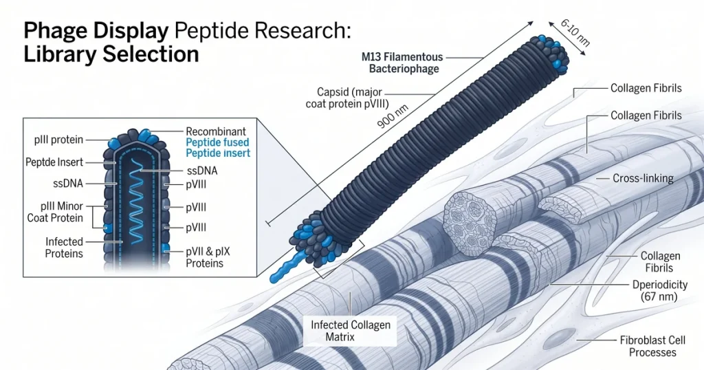
A phage display peptide library is a pool of M13 phages, each built to show a unique short peptide on coat protein pIII. A single pool can hold more than one billion distinct sequences for screening in one run. According to PubMed (2018), phage display helps find peptides with strong binding precision in isolated lab conditions.
Next Level Pharm provides research-grade, COA-confirmed peptides for these library testing workflows. Each vial is HPLC and mass spectrometry tested to a 99.4% average purity across the last 100 batches. A batch-specific certificate of analysis ships with every order.
Phage display relies on M13 as the primary vector because it replicates without lysing the bacterial host. This property allows large libraries to be grown and screened across multiple rounds. The method covers library construction, iterative panning, and synthetic hit validation.
Key Takeaways
- Genotype-Phenotype Link: Each phage connects its displayed peptide to the encoding gene inside the particle, enabling sequence recovery after selection.
- Library Diversity: A single M13-based library can hold over one billion unique peptide sequences for simultaneous screening against one target.
- Biopanning Cycle: Three to five rounds of binding, washing, and amplification enrich the library to isolate sequences with the highest binding affinity for the target.
- Adjustable Stringency: Wash conditions control how tightly a peptide must bind to survive each round, allowing labs to tune hit quality to their assay.
- Synthetic Testing: Confirmed hits are rebuilt as free peptides and retested by ELISA to check binding behavior outside the phage carrier format.
The sections below examine each workflow stage in sequence. Library construction, panning mechanics, stringency control, and hit validation are each covered in turn. This guide helps researchers design effective selection campaigns for specific binding targets.
What Is a Phage Display Peptide Library?
A phage display peptide library is a pool of M13 phages. Each phage shows one unique peptide on coat protein pIII. The peptide and its coding gene ride on the same phage particle.
This link between peptide and gene drives the panning process. Labs select phages by binding, then read the sequence from the gene. A single M13 library can hold over one billion unique sequences.
Library phages grow in a bacterial host after each panning round. The host amplifies only the phages that survived the wash step. According to Molecules (2020), M13 remains the most used format for peptide affinity screening.
A stable phage stock lets labs run three to five full panning cycles. Phages stay viable across all rounds when stored at the right conditions. Stock stays fit for use.
| Library Format | Peptide Length | Key Feature | Research Application |
|---|---|---|---|
| Linear random | 6-15 residues | Flexible chain | Broad initial screen |
| Constrained (disulfide) | 6-12 residues | Disulfide loop | Rigid receptor targets |
| Cyclic (head-to-tail) | 6-10 residues | Fixed backbone | Conformationally defined binding |
| D-amino acid | 6-12 residues | Protease-resistant | Stability-focused studies |
Format choice shapes the whole study. Linear types are most common.
How Are Libraries Constructed for Phage Display?
Library building starts by inserting random DNA into coat protein gene pIII. Each insert codes for a peptide of six to fifteen amino acids. Mixed codons at key spots create more range.
The total number of unique inserts sets the search range for panning. More range raises the chance of finding a strong binder. A standard M13 library holds over one billion unique sequences.
Molecular cloning checks that each phage holds one unique insert. This step blocks sequence overlap within the pool. The library is counted in culture before the first panning round.
Each stock is grown to a set titer before use. A defined titer confirms the right phage count enters each round. Good titer means good runs.
What Steps Make Up the Biopanning Cycle?
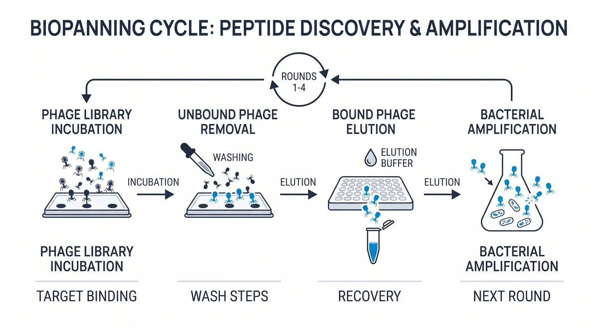
The biopanning cycle exposes the phage library to a target on a plate or bead. Phages that bind the target are captured on the surface. Unbound phages are flushed away by washing.
Bound phages are released by elution and grown in a bacterial host. This forms a new pool for the next panning round. Each round narrows the pool toward stronger binders.
The wash step is the key control point in each cycle. Buffer, detergent, and wash time all affect how many phages pass. By round four, the pool often holds a small set of tight binders.
Phage count drops with each round as weak binders are lost. This is the expected result of good pressure control. Count drops show clean work.
Browse research-grade peptides for validation work
How Is Selection Stringency Adjusted During Panning?
Selection pressure is the force a phage must hold to pass the wash step. Labs raise it by adding detergent, more salt, or longer wash time. Higher pressure removes phages with weak binding.
Longer wash time is one of the simplest ways to raise pressure. Extended washes flush out phages that are bound loosely. D-amino acid formats resist protease cleavage during wash steps.
Competitive elution adds a known ligand to displace only the tightest phages. Next Level Pharm stocks COA-confirmed peptide reagents for panning studies. According to NCBI PMC (2024), high-pressure selection improves binding precision in receptor assays.
Pressure is raised by one step per round in most designs. This gives the library time to respond at each level. This helps hits stay true.
How Are Hit Peptides Identified and Confirmed?
Hit peptides are found by sequencing phage clones from the final panning round. Each clone is grown alone to single-colony level. Its pIII insert is then read to reveal the peptide sequence.
Labs run ELISA on each clone to check for target binding. Clones that pass ELISA are rebuilt as free peptides via solid-phase synthesis. Free peptides allow direct binding tests without the phage carrier.
Peptide purity is checked by HPLC and mass spec on each batch. Next Level Pharm provides batch-specific COA records for all research compounds. Labs use these records to track purity across their selection campaigns.
Top-ranked hits move to further binding assays as research tools. A clear rank list helps labs select the lead candidate. Each hit gets a rank.
Frequently Asked Questions
What is the phage display selection cycle?
The selection cycle binds an M13 phage library to a target molecule fixed on a plate or bead. Phages that bind survive washing; non-binders are removed, and survivors are eluted and grown in bacteria for the next round. Each round enriches the library for stronger binders until a dominant set of high-affinity sequences emerges, and three to five rounds typically suffice.
How is a peptide phage display library constructed?
Library construction inserts randomized DNA into phage coat protein gene pIII, with each segment coding for a peptide of six to fifteen amino acids. Molecular cloning confirms each phage carries one unique insert, and the library is titered in culture to verify phage count before panning. These steps set the diversity ceiling and batch parameters for the entire selection campaign.
What selection pressure is applied in phage display panning?
Selection pressure is the stringency of wash conditions applied to remove phages with weak binding affinity at each panning round. Labs raise it by adding detergent, increasing salt levels, or extending wash time to push survivors toward stronger binders. High and even pressure across all rounds improves binding quality and the precision of all hits that reach the final selection cycle.
How are phage display hits validated after selection?
Confirmation begins by sequencing phage clones from the final selection round to read each displayed peptide sequence. Each confirmed clone is tested by ELISA to verify target binding before the sequence is resynthesized as a free peptide using solid-phase methods. The free peptide is confirmed pure by HPLC and mass spectrometry before use in downstream binding affinity assays and studies.
What peptide library formats are used in phage display research?
Common formats include linear libraries for broad screens and constrained libraries that use disulfide bonds to lock peptide shape for rigid receptor targets. Cyclic libraries add head-to-tail bonds for conformationally defined binding studies, while D-amino acid formats resist protease cleavage during wash steps. The choice of format depends on the target structure and the binding profile required for the assay.
What is the role of the M13 bacteriophage in phage display research?
M13 replicates without lysing the bacterial host, preserving the genotype-phenotype link as each library is amplified between all selection rounds. Its circular genome is easy to engineer with standard cloning tools, making high-diversity library construction practical across research settings. These core properties establish M13 as the standard phage vector for peptide display research in both academic and industrial laboratory settings.
How is biopanning used as the selection process in phage display?
Biopanning exposes a phage library to a target on solid support, removes non-binders by washing, and amplifies survivors in bacteria for the next round. This cycle repeats three to five times, and each round enriches the pool for sequences that bind more tightly to the target. After the final round, surviving phages are sequenced to identify the peptides that bound most consistently across all panning rounds.
What is the difference between phage display peptide and antibody display?
Peptide display presents short sequences of six to fifteen amino acids on phage coat protein pIII, while antibody display presents full antibody fragments. Peptide libraries are screened to find novel binding motifs and short linear peptides for target interaction and binding studies. Antibody libraries find high-affinity fragments that mimic natural immune recognition of specific protein targets in laboratory research contexts.
Summary
Phage display peptide library research relies on the M13 genotype-phenotype link. Each phage carries its coding gene inside and shows the peptide on its coat, letting biopanning reduce a billion-sequence library to a small confirmed set. Three to five panning rounds yield the top binders.
Key challenges include maintaining library diversity in early rounds and applying consistent stringency across all cycles. Synthetic testing with free peptide samples confirms that phage-based hits translate to the clean peptide format. Next Level Pharm supplies batch-tested, COA-verified research peptides for these validation assays.
What Should You Do Next?
Review your target molecule and select the library format that suits the binding study. Set wash buffer and detergent levels before the first round to ensure consistent selection pressure. COA-confirmed research peptides are available for all biopanning workflows.
Browse COA-verified research peptides
People Also Read
Peptide Microarray Research: High-Throughput Binding
Peptide Synthesis Explained: SPPS and Purity Standards
About the Author
Next Level Pharm Research Team
The Next Level Pharm research team is made up of biochemists and lab scientists. Their goal is to give researchers the highest-purity, COA-confirmed compounds available. Every batch is HPLC and mass spectrometry tested before dispatch.
Disclaimer: The information provided on this page is for educational and research purposes only. Next Level Pharm’s products are intended for laboratory research use only. They are not intended for human consumption, diagnostic, therapeutic, or medicinal purposes. This content does not constitute medical advice. Always consult a licensed healthcare professional before making any health-related decisions.