
Peptide-Lipid Membrane Interaction: Bilayer Research

Last updated: October 2026
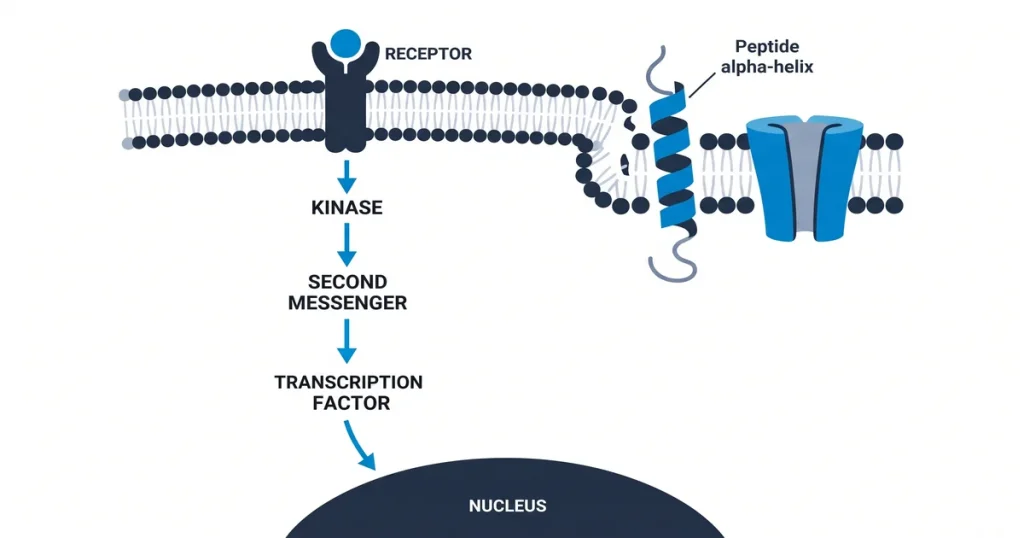
A peptide lipid membrane interaction is the study of how peptides bond to, enter, or cross the fat layers in cell walls. Labs watch how amino acid chains link to these lipids and learn how peptide shape affects cell entry. Data from the Journal of Peptide Science (2021) show that peptides bind well to charged lipids, making this a core topic for transport research.
Next Level Pharm provides high-purity research compounds for lab teams. The team holds a purity standard of 99% or higher for every lot. They average 99.4% purity across the last 100 batches.
Every vial goes through two tests: HPLC and mass spec. These two methods confirm what is in each vial. A COA (certificate of analysis) ships with every order.
third-party testing records is on their site. The team stores all stock in the USA. Average ship time is about 48 hours.
These compounds ship in lyophilized (freeze-dried) form. They are sealed under inert gas. This keeps them stable at room temp and cuts the need for cold chain shipping.
By offering stable materials, the team supports precise testing in many lab models. These traits matter for labs that want to see how peptides act on synthetic membranes. The next sections cover the core science behind these processes.
Key Takeaways
- Core Roles in Biology: Peptide and lipid contacts control cell signals and transport. These steps form the basis of key defense roles in the body.
- Lab Model Systems: Scientists use synthetic membranes like liposomes to mimic cell settings. These models let teams watch membrane contacts up close.
- Key Lab Tools: Labs measure binding with tools like circular dichroism (CD) and calorimetry.
This process is vital to keep cells alive. It also manages the flow of chemical signals through the body.
Antimicrobial peptides (AMPs) are germ-fighting compounds. They often work by making pores in bacterial walls. This leads to cell death through loss of balance.
Other chains called cell-penetrating peptides (CPPs) carry cargo across walls without causing harm. A peptide’s role in signal transfer depends on how well it moves through the membrane. Proper protein folding in membranes relies on nearby lipids to hold the final shape.
What models simulate cell membranes for study?
Synthetic membrane systems mimic real cell walls in controlled settings. They copy the physical traits of natural bilayers. These models let labs see how peptides insert into, span, or break a membrane.
Langmuir monolayers let labs study how peptides line up at the air-water surface. These models are key for testing shape changes in a controlled setting. Tirzepatide is on hand as a COA-tested research peptide.
Every lot is tested by HPLC and mass spec. These systems make sure every reading stays precise and repeatable.
How are these interactions measured in labs?
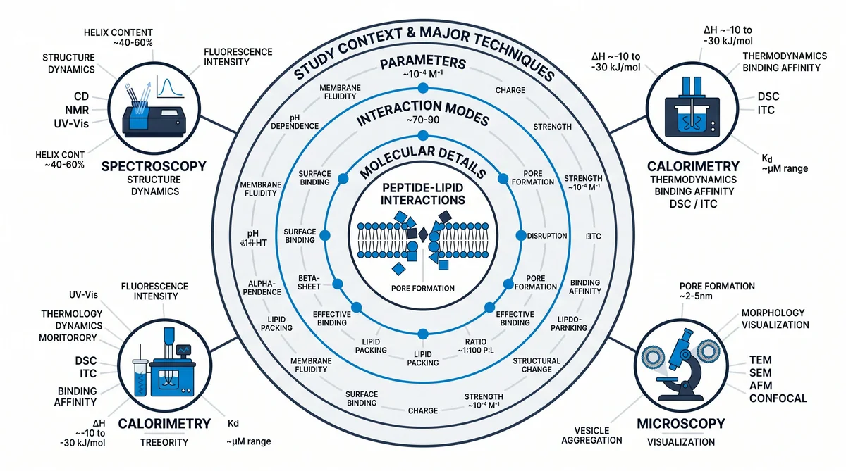
Labs measure these contacts with a set of physics-based tools. These methods track how peptides bind to and change lipid membranes.
CD spectroscopy tracks shifts in peptide shape when it meets lipids. ITC (isothermal titration calorimetry) measures the heat of binding. This maps the energy profile of the contact.
Fluorescence spectroscopy shows changes in the local setting of the peptide. This points to binding depth or membrane entry. AFM (atomic force microscopy) gives visual data on physical changes in the lipid layer.
By joining these tools, labs build a full dataset on membrane affinity. According to the Journal of Peptide Science (2021), these tools give high precision for tracking membrane-peptide contacts.
What peptide properties drive membrane binding?
A peptide’s pull toward a lipid membrane depends on its physical traits. These include its amino acid makeup and 3D shape. These traits set how it first touches and then enters the bilayer.
According to the Journal of Peptide Science (2020), amino acid layout in a peptide is the main factor in binding strength.
Positive charge acts as the first signal. It pulls the peptide toward negative groups on the membrane surface. Water-repelling traits then push it into the lipid core.
Many membranes have a two-layer setup. So the peptide orients its non-polar parts toward the inside. This setup often leads to stable pores or channels across the barrier.
The shape of the peptide (such as an alpha-helix) sets how well it stays in the membrane. If it cannot form the right shape, the bond weakens. These factors work together to set the binding result.
What are the primary models of interaction?
The main models describe how peptides break or pass through a lipid bilayer. These include the barrel-stave, toroidal pore, and carpet models. Each one explains a distinct pattern of membrane contact.
Some contacts form brief pores that create short-lived channels. Labs tell these apart by watching the peptide’s angle next to the membrane. Synthetic bilayers help them map each mode of entry.
In the barrel-stave model, peptides push in at a right angle. They group to form a protein-lined pore, like staves in a barrel. The carpet method works in a different way.
Peptides pile up on the surface until they reach a high level. Then the membrane breaks apart. The toroidal pore model shows a third path where peptides cause lipid bending.
The membrane lipids curve to line the pore along with the peptide chains. According to Molecules (MDPI) (2021), these changes are key paths for peptide-driven membrane shifts. These models help labs sort how various sequences affect the lipid bilayer.
What are the research implications of this field?
Studies of peptide-lipid contacts focus on building better transport tools. By learning how peptides link to membranes, scientists design molecules that boost delivery precision. These studies give the data needed to build fusion blockers that stop viral entry.
Also, control of these contacts supports the design of body-safe materials. These materials work with living systems without sparking an immune response. These gains form the basis for next-step work in molecular science.
Current studies also show these peptides can help build new antibiotic pipelines. Labs aim to make sequences that target bacterial walls but leave host cells alone. This focus meets the urgent need for tools that fight resistant bacteria in lab models.
By using the rules of membrane breakdown and selective binding, these designs raise the chance of targeted action. These results set a clear path for future work on membrane-active compounds.
Frequently Asked Questions
What is the mechanism of peptide-lipid bilayer interaction?
According to PubMed (2024), this topic draws strong research interest. Peptide-lipid contacts start when a peptide moves from water to the membrane surface. Positive charges pull it toward negative lipid heads. Then, water-repelling parts dive into the fatty core of the bilayer. According to Peptides (ScienceDirect) (2022), this step often causes shape changes that lock the peptide into the membrane.
How are model membrane systems prepared for peptide studies?
Labs often use thin film methods to build synthetic vesicles for membrane study. They dissolve lipids in a solvent, then dry it to leave a thin lipid film on the vessel wall. A water-based buffer (sometimes with peptide added) meets the film and starts the self-build of closed vesicles. According to Molecules (MDPI) (2020), steps like shaking or pressing then refine vesicle size for steady lab results.
What biophysical techniques measure peptide membrane insertion?
Labs use tryptophan glow quenching to find how deep a peptide sits in a bilayer. CD spectroscopy spots shape changes upon membrane contact. ITC measures the heat of binding to map energy profiles. AFM lets teams see surface shifts during entry events. According to Peptides (2021), these combined tools give a full view of how peptide entry reshapes the lipid bilayer.
How does peptide hydrophobicity affect membrane interaction?
Water repulsion is a key driver for peptide entry into lipid bilayers. Peptides rich in non-polar amino acids show a stronger pull toward the fatty lipid core than those with polar chains. As water repulsion goes up, the peptide moves deeper into the membrane. This link sets how well a peptide bonds to the lipid phase in controlled synthetic test settings.
What membrane disruption assays are used in peptide research?
Labs often use dye-leak tests to watch for loss of membrane strength after peptide contact. These tests trap dyes inside liposomes that glow brightly when the wall breaks open. Another method tracks marker release from synthetic vesicles with light tools. According to PubMed (2020), these tests let labs measure how much a peptide breaks model membranes at each level tested.
What is the difference between a liposome and a micelle in membrane research?
Liposomes are sphere-shaped sacs with a lipid bilayer that holds water inside. They closely mimic a real cell wall. Micelles are smaller round clusters where lipids group with their fatty tails inward and polar heads facing out. Labs pick liposomes to study membrane passage or peptide entry. They use micelles to give peptides a fat-like setting for shape studies in water.
What role do cell-penetrating peptides (CPPs) play in research?
Cell-penetrating peptides are special sequences studied for their skill at crossing cell walls without lasting harm. Labs look at them as carriers that move cargo into cells. Research shows they enter through paths like direct crossing or uptake by the cell. Studies keep probing how the positive charge of these peptides lets them cross lipid barriers in controlled lab models.
How does membrane fluidity influence peptide binding events?
Membrane fluidity refers to how freely lipids move side to side in the bilayer. Cholesterol and fat type often control this trait. Higher fluidity speeds up lipid motion and can pull peptides to the surface faster. Rigid, ordered bilayers raise the energy cost for peptide entry. So the physical state of the lipid layer controls binding speed and peptide angle.
Are there computational methods to study peptide-lipid interactions?
Computer studies use molecular dynamics (MD) to model how peptides act with lipid bilayers atom by atom. These programs predict peptide angle, depth, and shape shifts in real time. Scientists use them to see events that are hard to capture with lab tools alone. According to Frontiers (2023), these digital models pair well with lab data for a close-up view.
What is the difference between the barrel-stave and toroidal pore interaction models?
In the barrel-stave model, peptides insert so their water-repelling sides face the lipids. Their water-loving sides then form the inner pore opening. In the toroidal pore model, the peptide makes lipid heads bend and curve to line the pore wall along with the peptide itself. According to Frontiers (2021), the lipid makeup and peptide shape together decide which model applies in a given membrane setting.
Summary
Check your study design to confirm peptide levels and liposome needs for your next lab tests. Match your data needs to the lot-specific COA for your chosen compounds. These documents hold the exact purity results for your stock and confirm supplies meet the 99% or higher threshold.
Next Level Pharm provides high-purity research materials for labs that value data accuracy. Browse the Shop for research peptide products.
What Should You Do Next?
Confirm that your study design specifies the peptide concentration, purity threshold, and membrane model system for each assay. Review the lot-specific Certificate of Analysis for each compound to verify purity meets the ≥99% standard.
Next Level Pharm stocks COA-verified research compounds for lab teams that need consistent purity across batches. Browse research peptides to source materials for your next study.
Browse recovery peptides for COA-verified research compounds. Browse fitness peptides for COA-verified research compounds.
People Also Read
- Liposomal Peptide Encapsulation Research: Bilayer Studies
- Self-Assembling Peptide Nanofiber Research: Scaffold
About the Author
Next Level Pharm Research Team
The Next Level Pharm research team is made up of biochemists and lab scientists. Their goal is to give researchers the highest-purity, COA-tested compounds on the market. Every batch is HPLC and mass spec confirmed before dispatch.
Disclaimer: The information provided on this page is for educational and research purposes only. Next Level Pharm’s products are intended for laboratory research use only. They are not intended for human consumption, diagnostic, therapeutic, or medicinal purposes. This content does not constitute medical advice. Always consult a licensed healthcare professional before making any health-related decisions.