
Liposomal Peptide Encapsulation Research: Bilayer Studies

Last updated: October 2026
A liposomal peptide encapsulation research is the study of how lipid vesicles protect and carry peptide compounds in a lab setting. These vesicles form a bilayer shell around a water core that guards the cargo from enzymes and pH shifts. Studies show encapsulation yields of 50 to 85 percent, making them a standard tool for peptide delivery research.
Next Level Pharm supplies lab-tested research peptides for delivery studies, with an average purity of 99.4 percent across its last 100 batches. Each vial is verified by HPLC and mass spectrometry before dispatch. A COA ships with every order and third-party testing records is available online.
Liposomes serve as model carriers in peptide delivery research. They can hold both water-loving and fat-loving compounds. This dual trait makes them useful across a wide range of peptide types.
Key Takeaways
- Liposome Bilayer Structure: A liposome is a sphere made from a phospholipid bilayer around a water core.
- Peptide Protection Mechanism: The bilayer shields peptides from enzymes and pH stress during the study period.
- Standard Preparation Methods: Labs use thin-film hydration and extrusion to control vesicle size and batch quality.
- Encapsulation Efficiency Metrics: Labs separate free peptide by centrifugation, then measure it with UV absorbance.
- Stability Factors: Cholesterol content, surface charge, and storage pH all affect vesicle integrity over time.
These five factors guide how researchers build and check liposomal peptide batches. Size, charge, and EE are the first data points labs log before a study run begins. The sections below cover each factor with the core data and tools in use today.
What are liposomes in peptide research?
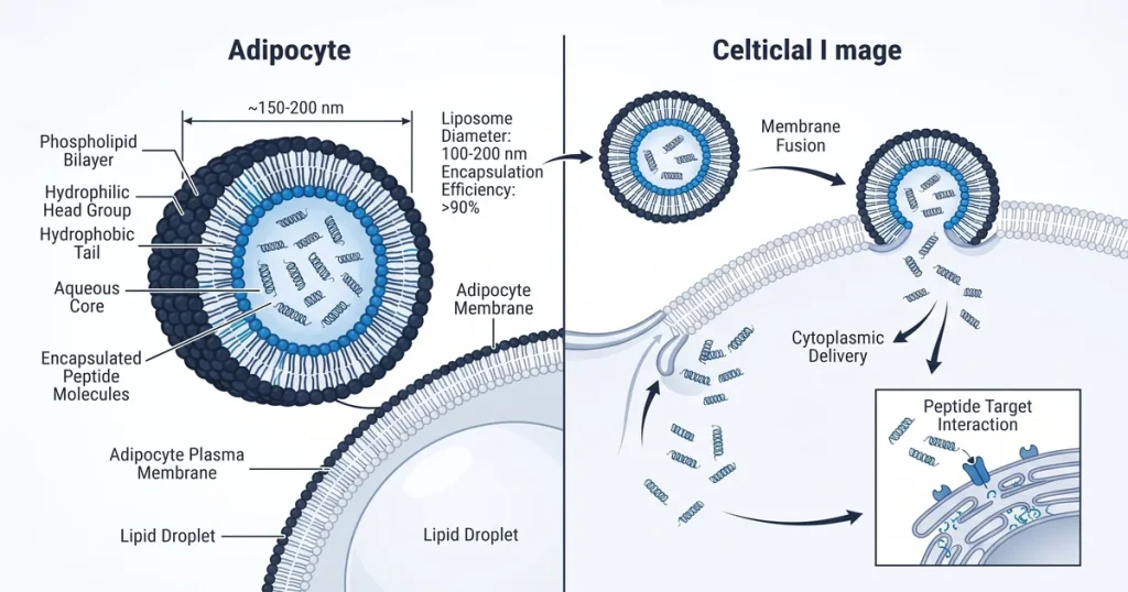
A liposome is a microscopic sphere built from one or more phospholipid bilayers around a water core, used as a carrier in peptide research. Phospholipid molecules have a water-loving head and a fat-loving tail. In water they form a closed shell that traps a small water volume inside.
Researchers use this shell to isolate peptides from the outer medium. Water-loving peptides dissolve in the inner core. Fat-loving peptides embed in the bilayer wall.
This dual trait suits a wide range of test compounds. GHK-Cu and BPC-157 are two peptides often studied in liposomal delivery work at Next Level Pharm.
The bilayer wall is about 4 nm thick. This thin shell is strong enough to keep the inner water core apart from the outer buffer. Labs rely on this property for tests that run for hours or days without re-dosing the well.
Vesicle size ranges from 80 to 200 nm in most lab work. This size lets the vesicle move through a cell medium by normal uptake routes. Labs pick the size to match the cell model they are using.
How does encapsulation shield peptide cargo?
Encapsulation shields peptide cargo by placing a lipid membrane between the molecule and the agents in the outer solution. Enzymes called proteases cannot cross the intact bilayer. So the peptide chain stays whole for longer in the test medium.
According to PubMed (2019), liposomal encapsulation raises the half-life of short peptides in cell media by two to four times. The shell also buffers the inner core from pH swings. Stable inner conditions help keep the sequence intact for the full study period.
The inner pH stays near the buffer set at fill time. This matters for peptides that break down fast in acid or base. Labs set the fill buffer between pH 6.5 and 7.4 to match the test model.
What preparation methods do labs study?
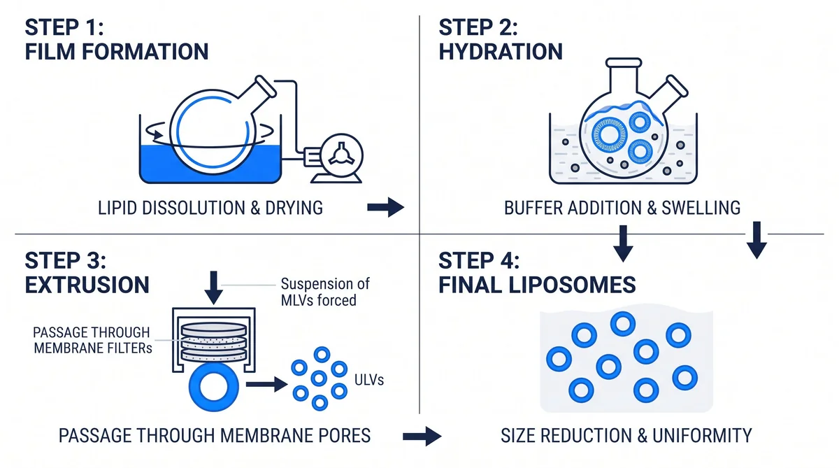
Labs use thin-film hydration as the main way to make liposomes. The process starts with lipids dissolved in a solvent. The solvent is then removed under low pressure to leave a dry film.
Adding a water buffer to the film causes the lipids to swell and form vesicles. Labs push the vesicles through a pore filter to set a target size. According to Molecules (MDPI) (2020), this extrusion step gives a narrow size range that improves batch consistency.
Sonication offers a faster route by breaking large vesicles into small ones with sound. Labs sourcing research peptides for delivery studies can browse the catalog at Next Level Pharm.
Freeze-drying the finished batch is a common way to extend storage life. The dry powder holds its structure at room temperature and is easy to ship. Labs reconstitute it in fresh buffer just before the run starts.
Batch size in most lab studies ranges from 1 to 50 mL. Small batches cut material cost and are easy to scale for pilot work. Labs run a test batch first to confirm the size and EE data before a full run.
How is encapsulation efficiency measured?
Encapsulation efficiency (EE) is the share of peptide trapped in the vesicles out of the total amount added at the start. Labs first separate the trapped fraction from the free peptide in the outer solution. This step is needed to get clean data.
High-speed spinning is the most common way to do this. The spin pulls the heavy vesicles down and leaves the free peptide in the top layer. Labs then dissolve the pellet and read the peptide level by UV absorbance or HPLC.
Labs can also use gel columns to keep the free peptide apart from the trapped fraction. The column holds back small free-peptide molecules while the vesicles pass through. According to Molecules (MDPI) (2024), this column method avoids heat stress from the high-speed spin.
| Parameter | Typical Range | Tool |
|---|---|---|
| Encapsulation Efficiency | 50-85% | UV absorbance |
| Vesicle Diameter | 80-200 nm | Dynamic light scattering |
| Polydispersity Index | 0.05-0.25 | DLS |
| Surface Charge | -30 to -20 mV | Electrophoretic assay |
| Storage Temperature | 2-8 degrees C | Stability log |
What factors affect liposome stability?
Liposome stability depends on lipid type, cholesterol level, surface charge, and storage pH. These four traits set how long a vesicle holds together without leaking. Lipid type sets baseline rigidity, while cholesterol fills bilayer gaps to raise it further.
Surface charge is key for keeping vesicles apart in a solution. According to Peptides (ScienceDirect) (2021), values below -25 mV or above +25 mV signal strong repulsion between particles. This repulsion stops aggregation in the suspension.
Cold storage at 2 to 8 degrees C reduces membrane stress and extends shelf life in most lab protocols.
Labs also track leak rate over time using a dye-release assay. A low leak rate shows the batch will hold for the full study. Most batches stay stable for 30 days at 4 degrees C when stored correctly.
What are the main research applications?
Liposomal peptide systems are used in lab research to study cell uptake, paths inside cells, and slow release of cargo. Labs label the outer surface with a dye to track vesicle movement in real time. The dye shows when and how the vesicle enters a target cell.
- Cell uptake studies map endocytosis paths in culture models.
- Slow release profiles measure how long the peptide persists in cell media.
- Surface tests add ligands to the outer wall to target specific cell lines.
- Fusion models study how vesicles merge with cell membranes.
According to PubMed (2022), surface-modified vesicles show higher uptake rates in epithelial cell lines than plain ones do.
Frequently Asked Questions
What is liposome encapsulation efficiency for peptides?
Encapsulation efficiency (EE) is the share of peptide trapped inside the vesicles out of the total amount put into the batch. Labs measure it by comparing the pellet peptide mass to the starting amount, with results varying by charge, size, and solubility. According to Peptides (ScienceDirect) (2021), EE ranges from 50 to 85 percent for small amphiphilic peptides in standard batches.
How are liposomes prepared for peptide encapsulation?
Thin-film hydration is the most widely used method in liposomal peptide research. Labs dissolve lipids in solvent, remove the solvent to form a dry film, then add a peptide buffer to form vesicles.
According to Molecules (MDPI) (2020), the vesicles are then pushed through a pore filter to set a narrow size range. This step improves batch uniformity from run to run.
What lipid types are used in peptide liposome research?
Phosphatidylcholine (PC) is the most widely used lipid in research batches. Labs blend it with cholesterol and charged lipids to tune the membrane traits needed for each test. Saturated lipids produce a more rigid bilayer with lower peptide permeability than unsaturated types.
According to Peptides (ScienceDirect) (2021), lipid choice directly sets how fast the trapped peptide diffuses out of the vesicle over time.
How is liposome size and polydispersity measured?
Dynamic light scattering (DLS) is the main tool for measuring vesicle diameter and polydispersity index (PDI), which shows how uniform the batch size is. A low PDI below 0.2 signals a narrow, well-controlled size range. Electron microscopy gives a direct visual check of shape and surface, and labs use both tools together to verify batch quality before the study starts.
What release kinetics do liposomal peptides show in studies?
Liposomal peptide release follows a two-phase pattern in most lab studies with standard phospholipid vesicles. The first phase is a fast burst as surface peptide dissolves into the medium. A slower phase then follows, driven by membrane wear and erosion over time.
According to PubMed (2022), the burst-to-slow ratio depends on how much peptide sticks to the outer bilayer surface during batch prep.
What is the difference between liposomes and niosomes?
Liposomes use natural phospholipids as the bilayer wall, while niosomes use synthetic non-ionic surfactants as the outer shell. Both form closed vesicles that hold water-loving cargo in their core. Niosomes resist hydrolysis better because synthetic surfactants are more stable in solution over time.
According to Molecules (MDPI) (2020), niosomes are favored when long storage stability is a high priority in peptide delivery studies.
How does surface charge affect liposome behavior in research?
Surface charge sets how liposomes interact with cell membranes and other particles in solution, with cationic vesicles binding more readily to negatively charged cell surfaces. Anionic vesicles resist non-specific binding, and neutral charge cuts clumping and sticking in salt-heavy buffers. Labs adjust the surface charge by picking specific lipids such as DOTAP for cationic or DOPS for anionic research batches.
Can liposomal peptides be used in cell uptake studies?
Yes, liposomal peptides are a common tool in cell culture work for studying how vesicles enter cells and release cargo inside them. Labs label the outer bilayer with a fluorescent dye to track vesicle movement and fusion events under a confocal microscope. According to PubMed (2022), this fusion path delivers higher peptide levels inside the cell than free peptide solutions do in standard culture.
Summary
Liposomal peptide encapsulation research gives labs a clear way to study how lipid shells protect and carry peptide compounds. The bilayer guards the peptide from enzymes and pH stress in the outer medium.
Key methods are thin-film hydration, extrusion, and sonication. Labs measure EE, vesicle size, and surface charge to check batch quality. Stability depends on lipid type, cholesterol level, and storage conditions.
Research uses span cell uptake work, slow release modeling, and surface modification studies. These data support more precise delivery systems in preclinical research. Labs that use COA-verified, high-purity peptides get a cleaner baseline for these models from the first run.
What Should You Do Next?
Review current lab protocols to confirm that lipid handling and storage match the batch specs for each compound. Confirm that storage temperature, fill buffer, and lipid-to-peptide ratio are logged for each batch. Check the COA for each lot before starting a formulation run.
Researchers who need lab-tested peptides for delivery studies can browse the full catalog at Next Level Pharm.
People Also Read
- Transdermal Peptide Delivery Research: Skin Permeability
- MAPK/ERK Pathway Peptide Research: Kinase Cascade
About the Author
Next Level Pharm Research Team
The Next Level Pharm research team is composed of biochemists and laboratory scientists dedicated to providing researchers with the highest-purity, COA-tested peptides available. Every batch is HPLC and mass spec confirmed before dispatch.
Disclaimer: The information provided on this page is for educational and research purposes only. Next Level Pharm’s products are intended for laboratory research use only. They are not intended for human consumption, diagnostic, therapeutic, or medicinal purposes. This content does not constitute medical advice. Always consult a licensed healthcare professional before making any health-related decisions.