
PPAR Gamma Pathway Peptide Research: Nuclear Receptor

Last Updated: October 2026
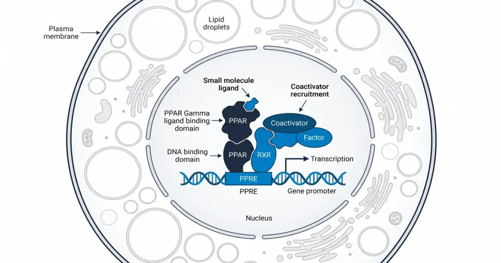
PPAR gamma pathway peptide research is the study of how synthetic compounds interact with PPAR gamma (peroxisome proliferator-activated receptor gamma, a nuclear receptor protein that regulates gene transcription). This receptor belongs to the nuclear receptor superfamily, a family of more than 48 transcription factor proteins. It governs gene programs in adipocytes (fat cells) and other metabolic tissues. According to PubMed (2024), covalent and non-covalent ligands can bind the same LBD pocket through distinct mechanisms, shifting the receptor between active and repressive states.
Next Level Pharm supplies research-grade compounds for in vitro nuclear receptor studies. The lab reports an average purity of 99.4% across the last 100 batches. Every vial is HPLC and mass spec verified before dispatch. Compounds ship lyophilized and stable at room temperature. COA (certificate of analysis, a document verifying purity and lot identity) documents accompany every order.
These materials support researchers working on nuclear receptor signaling. High purity reduces batch-to-batch variation in binding assays. Consistent samples make it easier to compare results across multiple cell lines. This page covers the key structures, tools, and methods used in PPAR gamma research.
Nuclear receptor research depends on understanding how ligands enter the binding pocket and how binding shifts the receptor conformation. PPAR gamma differs from other nuclear receptors by having a large, flexible LBD. This flexibility allows it to bind a wide range of molecules. Scientists use peptides to probe specific sites within this cavity.
Key Takeaways
1. PPAR Gamma Structure: The receptor has a LBD (ligand-binding domain), a DBD (DNA-binding domain, the segment that contacts target gene sequences), and an AF-2 (activation function-2) helix that controls coactivator docking.
2. Peptide Interaction Sites: Synthetic peptides carrying the LXXLL motif (a short leucine-rich sequence that coactivator proteins use to bind nuclear receptors) interact with the AF-2 surface of the LBD.
3. Biased Agonism: Research shows that different PPAR gamma agonists recruit different coactivator sets, a property called biased signaling.
4. Reporter Assays: PPRE-luciferase assays (PPRE stands for PPAR response element, a DNA target sequence that PPAR gamma binds) measure receptor activation in cell models.
5. Isoform Differences: PPAR gamma is found mainly in adipose tissue, while PPAR alpha (peroxisome proliferator-activated receptor alpha) is expressed mostly in the liver and oxidative metabolic organs.
6. Cell Line Selection: 3T3-L1 pre-adipocytes and HEK293 cells are standard models for PPAR gamma activation and gene expression studies.
Each of these points builds on established lab techniques. The sections below cover the receptor structure, peptide interaction methods, assay designs, and isoform comparisons. This reference supports researchers building protocols for nuclear receptor work.
What is the PPAR gamma nuclear receptor structure?
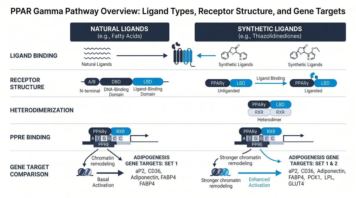
PPAR gamma is a nuclear transcription factor with four functional regions. These are the A/B domain, the C domain (DBD, the DNA-binding domain), the D hinge region, and the E/F domain (the LBD). The LBD forms a large hydrophobic pocket of roughly 1,300 cubic angstroms. This cavity allows diverse synthetic molecules to bind and shift receptor conformation. The AF-2 (activation function-2) helix sits at the C-terminal end of the LBD.
Ligand binding repositions the AF-2 helix. When the AF-2 is in an active position, coactivator proteins dock onto the receptor surface. When it is in a repressive position, corepressor proteins bind instead. According to PubMed (2024), covalent inhibitors shift the LBD conformational ensemble toward the repressive state by occupying the orthosteric pocket. This structural mechanism is central to all PPAR gamma peptide research.
How do peptides interact with the PPAR gamma ligand domain?
Peptides interact with the PPAR gamma LBD at two main sites. The first is the orthosteric pocket, where natural ligands like fatty acids bind. The second is the AF-2 coactivator surface, where LXXLL motif peptides make contact. Both sites are active targets in lab studies. This two-site model helps researchers test partial agonists alongside full activators.
LXXLL motif peptides bind the AF-2 surface directly. Changing residues around the core leucine sequence alters binding affinity. According to PubMed (2007), LXXLL-based peptides can block coactivator recruitment at the PPAR nuclear receptor interaction surface. This provides a tool for mapping which coactivators each ligand recruits. Phage display studies have identified optimized LXXLL variants for PPAR gamma research.
What reporter assays measure PPAR gamma activation?
Luciferase reporter assays are the primary method for measuring PPAR gamma activation. A PPRE-luciferase plasmid is cotransfected with a PPAR gamma expression vector in cell models. When PPAR gamma binds and activates the PPRE, the luciferase gene produces light. Luminescence levels reflect activation strength. This method maps dose-response relationships between test compounds and receptor activity.
TR-FRET (time-resolved fluorescence resonance energy transfer, a proximity-based detection method) is also widely used in nuclear receptor studies. It measures the interaction between the LBD and labeled coactivator peptides. When a ligand activates the receptor, the coactivator peptide moves close to the LBD surface. This generates a measurable signal. According to PubMed (2024), TR-FRET with NCoR1 (nuclear receptor corepressor 1, a protein that binds receptors in the inactive state) peptide probes can detect both activation and repression states in one assay.
How does PPAR gamma differ from PPAR alpha in signaling?
PPAR gamma and PPAR alpha share the same two-step nuclear receptor mechanism. Both heterodimerize with RXR (retinoid X receptor, a nuclear receptor partner required for DNA binding) and bind PPRE sequences in target gene promoters. However, they differ in tissue expression, ligand preference, and downstream gene targets. PPAR gamma is expressed mainly in adipose tissue. PPAR alpha is expressed mainly in the liver, kidneys, and cardiac muscle.
Ligand preferences also differ between the two isoforms. PPAR alpha responds to long-chain fatty acids and fibrate compounds. PPAR gamma responds to polyunsaturated fatty acids and lipid-derived molecules. According to PMC (2024), epigenetic mechanisms regulate both receptors, but they target different gene sets via distinct coactivator complexes. A compound active on PPAR alpha will not necessarily activate PPAR gamma pathways in the same cell model.
| Feature | PPAR Gamma | PPAR Alpha | PPAR Delta |
|---|---|---|---|
| Primary tissue | Adipose tissue | Liver, kidneys | Skeletal muscle, heart |
| Ligand type | Polyunsaturated fatty acids | Long-chain fatty acids, fibrates | Fatty acids, prostacyclin |
| Key gene targets | FABP4, ADIPOQ, CD36 | CPT1, ACOX1, PDK4 | ANGPTL4, ABCA1 |
| Standard cell line | 3T3-L1, HEK293 | HepG2, primary hepatocytes | C2C12 myotubes |
| Signal role | Adipogenesis, lipid storage | Fatty acid oxidation | Energy expenditure |
What cell lines are used for nuclear receptor research?
Several cell lines are standard in PPAR gamma nuclear receptor research. Model choice depends on the study goal. Some lines express PPAR gamma naturally. Others require transient transfection of PPAR gamma plasmids. Cell line choice affects how clearly researchers can detect receptor activity signals.
Key cell lines used in PPAR gamma studies:
- 3T3-L1: A mouse pre-adipocyte cell line. It expresses high PPAR gamma during differentiation. This is the primary model for adipogenesis research.
- HEK293: A human embryonic kidney cell line. Often transfected with PPAR gamma for reporter assays. It has minimal endogenous nuclear receptor background.
- RAW264.7: A mouse macrophage cell line. Used for PPAR gamma research in immune signaling contexts.
- Cos-7: A kidney fibroblast cell line. Used for cotransfection studies with PPAR gamma and RXR pairs.
- HepG2: A human hepatocyte-derived cell line. Useful for comparing PPAR alpha and PPAR gamma in the same metabolic context.
Research labs often compare results across two or more cell lines. This confirms that findings are not cell-type specific. Western blot verification of PPAR gamma expression is standard before starting reporter assays.
How do AICAR and metabolic peptides support PPAR gamma studies?
AICAR (5-aminoimidazole-4-carboxamide ribonucleotide, a synthetic nucleoside analog that activates AMPK) is used in cross-pathway nuclear receptor research. AMPK (AMP-activated protein kinase, a key energy-sensing enzyme) activation by AICAR can affect downstream PPAR gamma gene expression in adipocyte models. This makes AICAR a useful tool for mapping the interface between energy sensing and nuclear receptor programs.
Research-grade AICAR is available at Next Level Pharm. Each lot is COA-verified and HPLC-tested. Metabolic compounds like 5-amino-1MQ (a methylated quinoline compound that affects NNMT activity, a key metabolic enzyme) are also studied in pathway research. These tools let researchers probe pathways that interact with PPAR gamma gene programs. Lab-grade 5-amino-1MQ is available at Next Level Pharm. Cross-pathway studies build a fuller map of nuclear receptor links to energy metabolism in cell models.
Frequently Asked Questions
What is the PPAR gamma nuclear receptor structure?
PPAR gamma is a nuclear transcription factor with four structural domains. These include the ligand-binding domain (LBD) and the DNA-binding domain (DBD). An AF-2 activation helix at the C-terminal end of the LBD controls coactivator recruitment. The LBD forms a large binding pocket for diverse molecules. According to PubMed (2024), the AF-2 helix position determines whether coactivators or corepressors bind the receptor surface after ligand entry.
How do peptides interact with the PPAR gamma ligand domain?
Peptides interact with the PPAR gamma LBD at the orthosteric binding pocket and the AF-2 coactivator surface. LXXLL motif peptides (leucine-rich sequences found in coactivator proteins) bind the AF-2 helix region directly. Binding strength depends on residues flanking the core LXXLL sequence. Phage display studies have identified high-affinity LXXLL variants for PPAR gamma laboratory research.
What reporter assays measure PPAR gamma activation?
PPRE-luciferase assays are the standard format for measuring PPAR gamma activation in cell models. A PPRE vector and PPAR gamma expression plasmid are cotransfected before ligand addition. Ligand binding triggers luciferase production and a measurable light signal. TR-FRET assays measure direct LBD-to-coactivator peptide interaction. Both methods give quantitative data on receptor activation levels in controlled cell systems.
How does PPAR gamma differ from PPAR alpha in signaling?
PPAR gamma is expressed mainly in adipose tissue. PPAR alpha is expressed mainly in the liver and oxidative tissues. Both bind PPRE sequences as RXR heterodimers. However, they differ in ligand selectivity, coactivator preference, and downstream gene targets. According to PMC (2024), both receptors are subject to distinct epigenetic regulation patterns in metabolic cell models.
What cell lines are used for nuclear receptor research?
3T3-L1 cells are the primary model for PPAR gamma adipogenesis research. HEK293 cells are used for transient transfection reporter assays. RAW264.7 cells support immune pathway studies. HepG2 cells allow comparison of PPAR alpha and PPAR gamma in the same metabolic setting. Each model has a different endogenous nuclear receptor expression profile that affects assay background signals.
What is the role of RXR in PPAR gamma signaling?
RXR (retinoid X receptor) must partner with PPAR gamma to bind DNA target sequences. The PPAR gamma-RXR heterodimer binds PPRE sequences in target gene promoters. Without RXR, PPAR gamma cannot effectively contact DNA. Researchers often transfect both PPAR gamma and RXR expression plasmids in reporter assay models. This ensures full pathway activity and clean signal detection in cell systems.
What is biased agonism in PPAR gamma research?
Biased agonism occurs when different ligands activate the same receptor but recruit different coactivator sets. In PPAR gamma studies, some partial agonists selectively recruit one coactivator over others. According to PMC (2024), both partial and full agonists of PPAR gamma can display biased signaling profiles. Researchers study this property using TR-FRET coactivator profiling in HEK293 or 3T3-L1 cell models.
How is PPAR gamma related to adipogenesis research?
PPAR gamma is a master regulator of adipogenesis (fat cell differentiation from pre-adipocyte precursors). Studies in 3T3-L1 cells show that PPAR gamma is required for cells to convert from pre-adipocytes to mature adipocytes. Researchers track PPAR gamma target genes like FABP4 (fatty acid binding protein 4) and ADIPOQ (adiponectin) to confirm differentiation. These gene markers confirm that receptor activation has occurred in the cell model.
What is the NCoR1 peptide used for in PPAR gamma assays?
NCoR1 (nuclear receptor corepressor 1) peptides are used in TR-FRET assays to detect receptor repression states. When a PPAR gamma ligand stabilizes the repressive conformation, NCoR1 peptide binding increases. This is the opposite of what coactivator peptide binding shows. Researchers use competing NCoR1 and coactivator peptide signals to classify compounds as agonists, partial agonists, or inverse agonists in binding assays.
What are PPAR response elements and why do they matter?
PPRE (peroxisome proliferator-activated receptor response element) sequences are the genomic targets of PPAR gamma. These short DNA sequences sit upstream of target genes in promoter regions. When the PPAR gamma-RXR heterodimer binds a PPRE, it activates transcription of that gene. Researchers place PPRE sequences upstream of reporter genes like luciferase to measure pathway activation directly in cell models.
Summary
PPAR gamma pathway peptide research maps the full cycle of nuclear receptor activation, from ligand entry into the LBD to coactivator recruitment and target gene expression. The receptor’s large, flexible binding pocket and prominent AF-2 helix make it a well-characterized model for nuclear receptor science. Tools like PPRE-luciferase reporters and TR-FRET coactivator assays give researchers quantitative data on each step. PPAR gamma differs from PPAR alpha in tissue distribution, ligand preferences, and coactivator profiles. Cell lines like 3T3-L1 and HEK293 provide standard platforms for testing these interactions in controlled settings.
What Should You Do Next?
Researchers planning PPAR gamma studies should confirm which reporter assay format fits their model. Use luciferase assays for gene-level data or TR-FRET for direct LBD interaction data. Select peptide tools with high purity to reduce false signals in binding assays. Verify PPAR gamma expression levels in your chosen cell line by Western blot before starting. Pair nuclear receptor compounds with validated COA documentation for traceable results.
Browse the full catalog of research-grade peptides and metabolic compounds at Next Level Pharm. Researchers studying fat metabolism pathways can also explore the weight loss peptide category for compounds used in adipocyte studies.
People Also Read
- AOD-9604 Research: What Studies Show About This HGH Fragment
- HGH Fragment 176-191 Research: Adipocyte Biology and Lipid Studies
About the Author
Next Level Pharm Research Team
The Next Level Pharm research team is composed of biochemists and laboratory scientists dedicated to providing researchers with the highest-purity, independently verified peptides available. Every batch is HPLC and mass spec verified before dispatch.
Disclaimer: The information provided on this page is for educational and research purposes only. Next Level Pharm’s products are intended for laboratory research use only. They are not intended for human consumption, diagnostic, therapeutic, or medicinal purposes. This content does not constitute medical advice. Always consult a licensed healthcare professional before making any health-related decisions.