
TGF-Beta SMAD Pathway Peptide Research: Signaling

Last Updated: October 2026
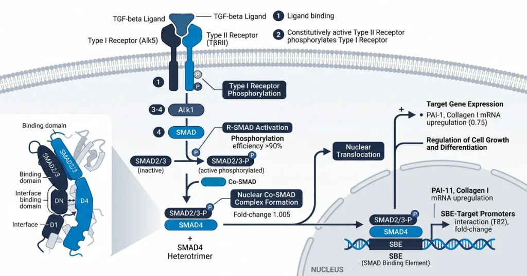
A TGF-beta SMAD signaling peptide research study is an investigation of how molecules affect the transforming growth factor beta pathway. TGF-beta signals through type I and type II serine-threonine kinase receptors. Receptor activation phosphorylates SMAD2 and SMAD3. These then complex with SMAD4 to enter the nucleus. Lab models use synthetic peptides to map each phosphorylation step and its effect on gene expression.
Next Level Pharm provides high-purity research compounds for researchers worldwide. The average purity across the last 100 batches is 99.4%. Every vial gets tested by HPLC and mass spec. All compounds are lyophilized and stored under inert gas. This keeps them stable at room temperature during shipping. No cold chain is needed. The lab holds over 70 active peptide SKUs with dispatch within 48 hours.
This focus on quality gives researchers consistent materials for testing TGF-beta pathways. Reliable peptide sequences map how signals flow from the cell membrane to the nucleus. These models show how SMAD proteins affect gene expression in cell lines. High standards lead to more accurate findings in cell signaling studies.
Key Takeaways
- TGF-beta Pathway Regulation: This signaling cascade controls core cell processes like growth, cell change, and programmed cell death.
- SMAD Protein Function: These cell-level transducers carry signals from cell surface receptors directly into the cell nucleus.
- Peptide control Potential: Research studies how specific peptides act as agonists or blockers to affect this TGF-beta pathway.
- Dual Signaling Cascades: Signaling occurs through canonical SMAD-dependent paths or non-canonical pathways like MAPK and PI3K/AKT.
- Standard lab Methods: Scientists analyze this pathway using Western blotting and luciferase gene activity assays.
These core elements set the framework for lab study of signal paths. The sections below cover specific mechanisms, control methods, and ways to track protein activity.
What is the TGF-beta signaling pathway?
The TGF-beta (transforming growth factor-beta) signaling pathway is a cell communication system. It regulates core processes like cell growth, tissue differentiation, and apoptosis. This pathway senses external cues to adjust cell behavior. According to the NIH (2020), this system is key for tissue stability and cell life cycles.
Signaling starts when a TGF-beta ligand binds to a type II receptor on the cell membrane. The type II receptor recruits and phosphorylates a type I receptor. This triggers a cascade through SMAD proteins to affect gene output. Balancing growth and cell death, this pathway is a key lab subject. Scientists examine these interactions to document how cell signals work in controlled models.
How do SMAD proteins mediate TGF-beta signals?
SMAD proteins mediate TGF-beta signals by acting as messengers inside the cell. They transfer external signals into the nucleus. The TGF-beta pathway relies on these proteins to convert binding events into gene changes. When the receptor is active, it starts a chain that shifts the cell state. This is the core way these signals alter cell function.
When a type I receptor is active, it adds phosphate groups to R-SMADs. These include SMAD2 and SMAD3. These proteins form a complex with SMAD4. The complex moves into the nucleus to regulate target gene output. Nuclear entry is a required step for the signal to work. SMAD2/3 is available as a COA-verified research peptide. Every lot is verified by HPLC and mass spec.
Which peptides can affect this pathway, which peptides can affect this pathway?
Some research peptides can interact with and affect the TGF-beta/SMAD cascade. These compounds let researchers probe tissue remodeling and fibrosis through lab steps. By targeting points in this cascade, researchers change how cells read signals for ECM making. The peptide GHK-Cu has shown effects on TGF-beta expression in tissue models. Researchers also use synthetic peptides as pathway agonists or antagonists. These either promote or block signaling. This lets labs observe fibrosis outcomes in test settings.
GHK-Cu is available as a COA-verified research peptide. Every lot is verified by HPLC and mass spec.
These agents help define the role of SMAD proteins in gene output. When researchers add an agonist, target gene transcription rises. An antagonist limits this. This control is key for studying how cells manage structure. Fibrosis studies track how proteins like collagen build up. By observing how peptides shift signaling balance, labs map each step in the cascade.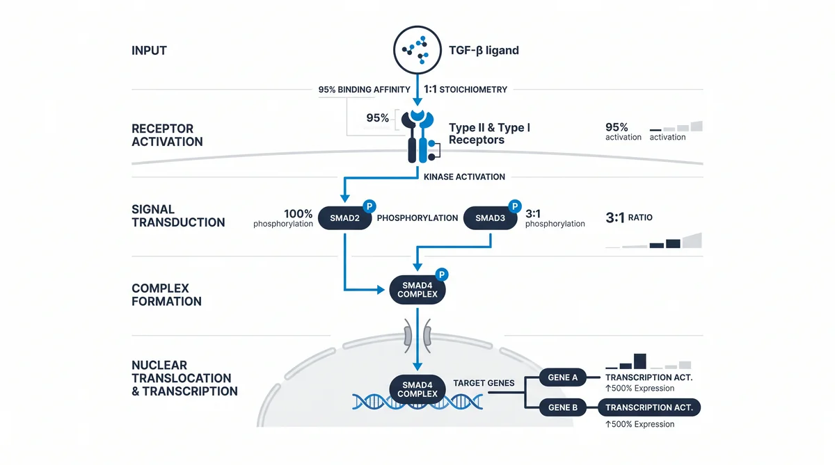
What are canonical vs. non-canonical pathways?
Canonical and non-canonical paths are the two main routes for TGF-beta signal flow. The canonical route uses SMAD proteins to start gene transcription through phosphorylation and cell entry. Non-canonical pathways work without SMAD and use other chains to alter cell behavior. Research shows these dual routes let ligands trigger a wide range of responses.
Non-canonical signaling often uses MAP kinase (MAPK) cascades and the PI3K/AKT pathway. These activate internal messengers outside the SMAD-dependent path. When a TGF-beta receptor binds its ligand, these cascades can affect cell structure or cell survival. Researchers study these routes to see how signals reach specific outcomes. According to Molecules (MDPI) (2018), crosstalk between these branches maps cell feedback loops.
How is this pathway studied in labs?
Researchers use several in vitro techniques to study the TGF-beta/SMAD pathway. These include cell culture models and biochemical assays. The methods track signal events in a controlled setting. Scientists use them to check how ligands or peptides affect SMAD phosphorylation. By analyzing these data, teams draw conclusions about cascade activity in various cell lines. These steps map how the signal flows from the cell membrane to the nucleus.
Common methods include Western blotting to detect SMAD phosphorylation and luciferase reporter assays to measure gene activity. RT-qPCR measures target gene changes. SMAD4 is available as a COA-verified research peptide. Every lot is verified by HPLC and mass spec. Researchers combine these tools to get a full picture of signaling shifts. Western blotting reveals protein levels. RT-qPCR confirms gene effects downstream. These combined methods ensure accurate findings on pathway control.
What are the research effects?
TGF-beta/SMAD research focuses on its dual role in oncology and fibrosis studies. Using peptides to alter this pathway, scientists study tissue repair and immune response. Lab models let teams isolate the effect of pathway binding on cell balance. These models show how selective blocking or stimulation alters disease over time. This is key for clarifying the switches that govern cell growth or programmed death.
Research peptides map SMAD signal dynamics in tissue settings. In fibrosis, they show how ECM buildup is controlled by TGF-beta. In oncology, the focus is how the pathway affects tumor environments. By adjusting receptor binding, teams gather data on immune response. These findings help build a clearer map of cell signaling in healthy and diseased states. Understanding these dynamics is key for tissue repair research.
Frequently Asked Questions
What is the TGF-beta SMAD signaling pathway?
This pathway transfers external signals into the cell nucleus. It controls gene output. Signals move from cell surface receptors through internal messengers. According to NCBI (2012), this framework is key for cell growth. It also governs tissue change and cell life cycles. It acts as a primary controller for many natural processes.
How do peptides affect SMAD phosphorylation?
Peptides influence SMAD phosphorylation by binding to cell surface receptors to mimic or block natural ligands. When these compounds interact with the TGF-beta receptor complex. They can shift the balance of protein activity within the cytoplasm. Research suggests that synthetic peptides can either accelerate or inhibit the ability of type I receptors to target SMAD proteins. lab assays monitor these effects to map how specific peptide structures change internal signaling intensity and duration.
What assays measure TGF-beta receptor activation in vitro?
Scientists measure receptor binding through Western blotting. This detects phosphorylated SMAD proteins in a cell sample. Luciferase reporter tests help researchers measure gene activity. According to the Journal of Peptide Science (2022), these tools give clear data on signaling. They allow precise tracking of pathway response in varied test conditions.
How do SMAD2 and SMAD3 differ in signal transduction?
SMAD2 and SMAD3 share structural traits. But they play unique roles in gene binding. SMAD3 often links directly to gene start sites. SMAD2 often needs extra proteins for DNA binding. Research shows these two proteins can have distinct cell effects. Scientists study these differences to see how cells generate specific responses.
What cell models are standard for TGF-beta research?
Standard models include HEK293 and other epithelial cell lines. These show clear TGF-beta responsiveness. They allow consistent tracking of signaling across tests. Researchers also use primary fibroblasts or stem cells. These reflect how the pathway works in specific tissues. According to NIH (2020), model choice is key for accurate signaling findings.
What is the function of SMAD4 in the pathway?
SMAD4 serves as the common mediator for both R-SMAD pathways. After SMAD2 or SMAD3 are phosphorylated, they form a complex with SMAD4. This complex must move into the nucleus. It then binds DNA to start gene output. Without SMAD4, the signaling chain cannot affect gene output. SMAD4 acts as the final gatekeeper for the signal.
What are blocking SMADs (I-SMADs)?
blocking SMADs, known as SMAD6 and SMAD7, function by preventing the activation of other SMAD proteins. They work by competing with R-SMADs for receptor binding or by triggering the breakdown of receptor complexes. Research suggests these proteins act as a negative feedback loop to ensure that signaling does not remain active for too long. By blocking the signal, I-SMADs help the cell maintain homeostasis. Prevent excessive growth or tissue remodeling responses.
Can TGF-beta signaling be inhibited for research?
TGF-beta signaling can be blocked using chemical blockers. These target the kinase activity of the receptors. Peptides that compete for ligand binding also work. These blockers let scientists isolate the pathway in tests. According to PubMed (2023), blocking this pathway is a standard research method. It helps find the role of TGF-beta in disease models.
What role does TGF-beta play in tissue fibrosis studies?
TGF-beta drives fibrotic processes by activating fibroblasts. It also promotes the making of ECM proteins. In lab models, researchers track how this pathway raises collagen buildup. This helps map the steps that lead to organ structural changes. Understanding these links is key for tissue repair research. It also helps map how scarring patterns form over time.
How is the TGF-beta SMAD pathway’s gene activity measured in a lab setting?
gene activity is measured by linking the activity of the pathway to a reporter gene that produces light or color. Researchers use a plasmid containing a promoter sequence that responds directly to SMAD complexes to measure the signal strength. This approach allows for a direct measurement of how much the pathway is influencing the cell at a specific moment. According to Molecules (2023), these assays provide a way to document the regulatory output of the entire signaling chain.
What Should You Do Next?
Review your current protocols to find gaps where peptides could clarify signaling pathways. Select peptides that match your study goals. Verify that your lab has equipment for storage and dilution. Once your objectives are set, visit the store to match your needs to catalog items. Review the COA for each lot to maintain baseline accuracy.
Researchers sourcing these peptides can explore the catalog at Shop. Every vial ships with a COA and full lot traceability.
People Also Read
- BPC-157 vs TB-500: A Detailed Research Comparison Guide
- GHK-Cu Research Explained: What Peer-Reviewed Studies Show
About the Author
Next Level Pharm Research Team
The Next Level Pharm research team is composed of biochemists and laboratory scientists dedicated to providing researchers with the highest-purity, independently verified peptides available. Every batch is HPLC and mass spec verified before dispatch.
Disclaimer: The information provided on this page is for educational and research purposes only. Next Level Pharm’s products are intended for laboratory research use only. They are not intended for human consumption, diagnostic, therapeutic, or medicinal purposes. This content does not constitute medical advice. Always consult a licensed healthcare professional before making any health-related decisions.