
CCK Peptide Research: Cholecystokinin Receptor Studies

Last Updated: October 2026
A CCK peptide research study is an investigation of how cholecystokinin, a 33-amino-acid hormone, signals through CCK1 and CCK2 receptor subtypes. CCK activates Gq protein-coupled pathways in pancreatic and neural tissue models. It is one of the first gut hormones identified, with a sulphated tyrosine residue critical for high-affinity CCK1 receptor binding. Lab models use synthetic CCK to probe these binding interactions.
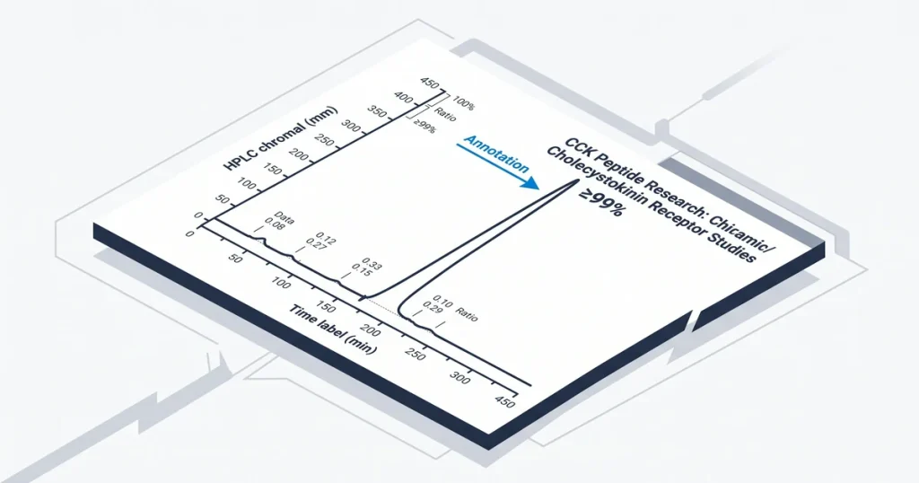
Next Level Pharm provides high-purity peptides for lab use. All compounds reach 99.4% average purity across the last 100 batches. Independent labs verify every lot by HPLC and mass spectrometry. All vials are stored in a lyophilized, inert atmosphere. This keeps them stable at room temperature with no cold chain required.
Labs monitor these pathways to refine energy control data. The CCK receptor system affects various tissues. Precise synthetic compounds allow for targeted study in controlled assays. Accurate molecular analysis requires materials that meet strict purity standards. Data from these trials depend on compound structural integrity.
Key Takeaways
- Core Biological Function: Cholecystokinin acts as a key peptide hormone. Neurotransmitter that helps regulate digestion and satiety.
- Primary Receptor Types: Research focuses on CCK-A. CCK-B receptors which have different tissue locations and binding affinities.
- Active Peptide Fragments: CCK exists in various forms. Researchers often select the sulfated octapeptide CCK-8 for lab studies.
- Intracellular Signaling Pathways: CCK receptors link to the Gq/11 pathway to increase calcium. Activate protein kinase C activity.
- Laboratory Research Antagonists: Selective agents like loxiglumide serve. As essential tools to isolate CCK pathways for specific observational analysis.
The primary CCK receptor subtypes define the scope of modern metabolic studies. These receptors dictate how chemical signals propagate through cell cascades during testing. Analyzing these mechanisms requires attention to ligand-receptor binding. Structural stability must be maintained across every sample.
What are CCK peptides and their primary functions?
Cholecystokinin (CCK) is a peptide hormone found in the GI tract. It also acts in the brain as a brain signal. CCK belongs to the cholecystokinin family of peptides. It is made by I-cells in the duodenum and jejunum. In these tissues, CCK acts as a chemical messenger for food arrival from the stomach. According to the NIH (2020), this peptide is vital for controlling gastric emptying and nutrient processing.
CCK stimulates gallbladder contraction to release bile. It also triggers pancreatic enzyme release into the small intestine. These actions help break down fats and proteins for nutrient uptake. Beyond digestion, CCK acts in the brain to signal satiety. By binding to specific receptors, it helps the body monitor energy intake. Cholecystokinin-8 is a COA-verified research peptide. Every lot is verified by HPLC and mass spec.
How do CCK-A and CCK-B receptors differ in studies?
CCK-A and CCK-B receptors differ mainly by their location. They also differ in their binding preferences for ligands. The CCK-A receptor stays concentrated in the GI system. It affects digestion. The CCK-B receptor is found in the brain. It is densely expressed there. These two proteins mediate separate pathways.
CCK-A receptors show strong binding for sulfated CCK peptides. This is their main trigger for signal. The CCK-B receptor binds both sulfated CCK and gastrin with high affinity. This allows the CCK-B type to respond to both peptides. CCK-8 Sulfated is a COA-verified research peptide. Every lot is verified by HPLC and mass spec. These distinct binding patterns lead to different natural responses in lab models.
What is the pathway of CCK receptor activation?
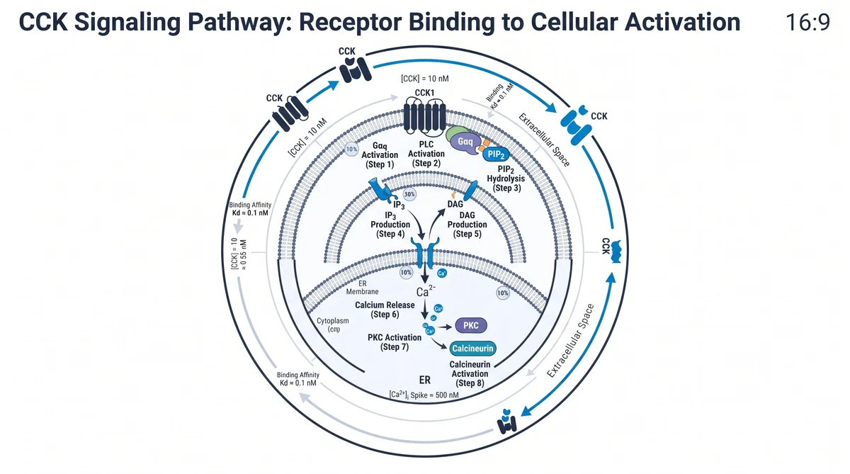
CCK receptors function as G-protein coupled receptors (GPCRs). They mainly couple to the Gq/11 protein. When a ligand binds, it causes a shape change that recruits Gq/11. This starts a signal cascade in the cell. According to PubMed (2002), this coupling allows precise control of cell responses in labs.
The Gq/11 protein triggers the enzyme phospholipase C (PLC). PLC breaks down PIP2 into two second messengers: IP3 and DAG. IP3 diffuses into the cytosol to release calcium from internal stores. This releases calcium inside the cell. DAG stays in the membrane and activates protein kinase C (PKC). These combined signals propagate the cell effects of CCK binding. CCK-8 is a COA-verified research peptide. Every lot is verified by HPLC and mass spec.
Which CCK fragments are studied most in research?
The most studied CCK fragments are CCK-8, CCK-33, and CCK-58. These arise through post-translational processing of a single pro-hormone. The parent molecule is processed in different tissues. Labs use these isoforms for isolated study. Sulfated CCK-8 is widely used due to its high natural potency. It captures the key signal profile for gut and brain responses, according to PubMed (1998).
CCK-8 is a COA-verified research peptide. Every lot is verified by HPLC and mass spec. CCK-8 is preferred for in vitro assays due to its simple structure. Research shows the sulfate group on the tyrosine residue is vital for high affinity at the CCK-A receptor.
What are CCK receptor antagonists used for in labs?
CCK receptor antagonists block cholecystokinin at its specific receptors. These agents let labs isolate and study CCK’s natural pathways. Labs apply these compounds in controlled settings. This helps determine how CCK affects natural signal without interference from other ligands.
Loxiglumide acts as a selective CCK-A antagonist. Proglumide blocks both receptor subtypes. These molecules help study CCK’s role in GI motility, pain response, and anxiety. Loxiglumide and Proglumide are available as COA-verified research peptides. Every lot is verified by HPLC and mass spec. These tools help labs see how receptor blockage alters cell output.
What future research directions exist for CCK?
Future CCK research involves novel receptor agonists. These explore impact on energy and brain pathways. Labs use these agents to study satiety pathways in animal models. The focus includes how these compounds interact with the brain. They may affect feeding behaviors.
CCK studies also extend to panic disorder models. Labs monitor receptor binding in specific brain regions. These studies examine if changes to signal cascades can alter anxious responses. Further work focuses on GI motility studies. Labs assess these peptides’ ability to influence gastric transit times. According to the NIH (2020), knowing these local effects helps map CCK’s role in the gut-brain axis.
CCK-8 is a COA-verified research peptide. Every lot is verified by HPLC and mass spec. Ongoing lab data support a broad scope for future work on peptide selectivity. Labs continue to improve these molecules to better understand their function.
Frequently Asked Questions
What is the CCKAR receptor structure in CCK research?
The CCKAR receptor belongs to the G-protein coupled receptor superfamily. Mapped by seven transmembrane alpha-helical domains. Its structure includes an extracellular N-terminal region that functions. As the primary binding site for the sulfated tyrosine residue of the peptide. Intracellular loops interact with G-proteins to initiate signal transduction. Scientists analyze these structural domains to understand how ligand binding triggers specific conformational changes. According to the NIH (2020).
How does CCK-8 differ from CCK-33 and CCK-58?
CCK exists as a family of peptides with different lengths. Including CCK-8, CCK-33, and CCK-58. These numbers refer to the count of amino acids within each specific chain. CCK-8 is the C-terminal octapeptide fragment that remains active in most biological assays, while larger forms circulate in the blood, the smaller CCK-8 fragment is frequently used in research to mimic the native activity of the hormone. According to the Journal of Peptide Science (2012).
What G-protein pathway does CCKAR activate?
Research shows that CCKAR primarily activates the Gq/11 signaling pathway. Once the peptide binds to the receptor, it triggers the activation of phospholipase C, which increases intracellular calcium levels and activates protein kinase C. This pathway serves as the main mechanism for CCK-mediated effects on cellular functions like enzyme release. According to PubMed (2002), the activation of these secondary messengers is a hallmark of CCKAR engagement during experimental finding.
How is cholecystokinin binding affinity measured?
Binding affinity is typically assessed using radioligand competition assays in membrane preparations. Researchers incubate the receptor-rich membranes with a labeled ligand. Then add increasing concentrations of the test compound to see if it displaces the original label. The concentration required to inhibit fifty percent of binding determines the affinity constant. This approach allows investigators to quantify how effectively a peptide interacts with the receptor. According to the NCBI (2007).
What in vitro models are used in CCK receptor studies?
Investigators use various cell lines, such as the AR42J pancreatic cell line or transfected CHO cells. To study receptor dynamics. These models provide a controlled environment to observe signal transduction, receptor internalization. Downstream gene expression without systemic variables. By isolating the receptors in these cell types, researchers can map the potency of different peptides with high precision. These standardized models remain essential for mapping signaling pathways. According to PubMed (1991).
Is CCK related to the peptide gastrin?
CCK and gastrin belong to the same family of peptides, because they share an identical C-terminal pentapeptide sequence. This structural similarity explains why they can exhibit overlapping activity at certain receptors in lab settings, while they share this common architecture, they originate from different gene expressions and fulfill distinct natural roles. Researchers carefully distinguish between these two to ensure the specificity of their findings. According to the NIH (2020).
What role does sulfation play in CCK activity?
Sulfation of the tyrosine residue at the seventh position from the C-terminus is critical for high-affinity binding to CCKAR. Research shows that non-sulfated forms exhibit notably lower potency. When interacting with these specific receptors. This chemical change acts as a biological switch that dictates the strength of the interaction within the gut and brain. According to the Journal of Peptide Science (2012), this structural feature remains a primary focus in synthesis and mapping studies.
Can CCK peptides cross the blood-brain barrier in lab models?
Evidence suggests that CCK peptides have limited ability to cross the blood-brain barrier through passive diffusion in their native forms. However, research into specific transport mechanisms or analog changes continues to be a point of study. Investigators monitor central nervous system effects by using specialized administration techniques or observing how peripheral signaling influences brain activity via the vagus nerve. According to the NIH (2020), these pathways are vital for knowledge systemic satiety.
What are CCK receptor antagonists used for in research?
Antagonists like loxiglumide are used to block receptor binding sites to isolate the contributions of natural CCK. These tools help researchers determine which natural responses rely on the activation of CCK-A or CCK-B receptors. By selectively shutting down these pathways, scientists can measure the baseline function of the hormone in digestion and satiety. This experimental control provides a clearer picture of peptide signaling. According to the NCBI (2010).
What is cholecystokinin’s role in satiety signaling in lab studies?
In preclinical models, administration of CCK is shown to reduce short-term food intake and promote satiety. The peptide acts as a signal to the brain. Often through the vagus nerve. To modulate feelings of fullness after a meal. Researchers use these models to study how the gut-brain axis monitors energy balance. According to PubMed (1989), these studies confirm that the peptide serves. As a significant satiety signal in animal research.
What Should You Do Next?
Audit your current lab inventory and confirm that your peptide storage protocols align with the structural requirements defined in your batch COA. Document the specific sequence verification results and lot numbers to maintain consistency across all ongoing experimental trials. You should also verify that your lab model allows for the precise isolation of CCK-A or CCK-B signaling pathways before starting new assays.
Next Level Pharm supports investigators by providing high-purity, independently verified compounds for their metabolic research needs. Researchers sourcing cholecystokinin or related signaling peptides can browse the catalog at Shop. Every vial ships with a COA and full lot traceability.
People Also Read
- What Are Nootropic Peptides? Mechanisms, Research Review
- What Is Selank? GABA Pathways, Mechanisms, and Study Findings
About the Author
Next Level Pharm Research Team
The Next Level Pharm research team is composed of biochemists and laboratory scientists dedicated to providing researchers with the highest-purity, independently verified peptides available. Every batch is HPLC and mass spec verified before dispatch.
Disclaimer: The information provided on this page is for educational and research purposes only. Next Level Pharm’s products are intended for laboratory research use only. They are not intended for human consumption, diagnostic, therapeutic, or medicinal purposes. This content does not constitute medical advice. Always consult a licensed healthcare professional before making any health-related decisions.