
PACAP Peptide Research: Adenylate Cyclase Studies

Last Updated: October 2026
A PACAP peptide is a neuropeptide in the secretin/glucagon superfamily. It potently stimulates the adenylate cyclase enzyme system. This peptide exists in two forms: PACAP-27 and PACAP-38. Research confirms this. Both forms interact with specific cell receptors. Scientists study these molecules to observe cell signaling. PACAP is a core study subject. Neural and endocrine research both use it.
Next Level Pharm supports these studies by providing high-purity research compounds for lab settings. Their protocols ensure 99.4% average purity. This holds across 100 batches. Every vial undergoes HPLC and mass spectrometry verification. This confirms chemical identity for each lot. The team keeps stock lyophilized and stable. It is sealed under an inert atmosphere. This approach gives researchers consistent materials.
These peptide samples ship from their USA-based facility. Average dispatch time is about 48 hours. A COA comes with every order. It covers each lot fully. Scientists use this data to confirm peptide profiles. This happens before each study. Verified purity helps labs maintain precise conditions. Records support each step.
Key Takeaways
- Potent Signaling Capability: This neuropeptide activates the adenylate cyclase enzyme to drive cellular signal transduction processes. It is a key step in cell signaling. Information passes within the cell.
- Distinct Bioactive Structures: It exists in two primary forms known as PACAP-38 and PACAP-27. These forms exhibit unique activities. Varying peptide lengths drive the differences.
- Specific Receptor Interaction: The PAC1 receptor maintains a high affinity and specificity for this peptide. This receptor sets PACAP apart from VIP. VIP binds to broader receptor targets.
- Neuroprotective Research Findings: Preclinical data show the peptide helps defend neurons against damage in lab settings. It activates anti-apoptotic and pro-survival pathways.
- Endocrine System Regulation: It acts as a primary modulator for the pituitary gland in natural models. Research shows it stimulates hormone release. Key endocrine hormones are affected.
The sections below explore the mechanisms of action. They cover receptor distribution in lab models. Researchers can review these details. They show how structure and binding affect outcomes. This applies to cell-based assays and tissue studies.
What is PACAP’s mechanism of action?
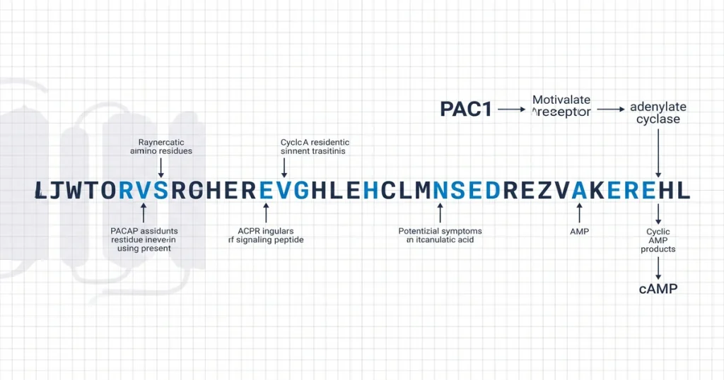
PACAP binds to G-protein coupled receptors. It mainly targets the PAC1 receptor. This starts complex signaling cascades inside cells. Once bound, PACAP modulates cell pathways. Biological responses are influenced. This interaction lets PACAP transmit signals. They cross cell membranes. According to the Journal of Peptide Science (2018), receptor binding activates downstream messengers. This was shown in lab models.
The main mechanism involves two pathways. The first is the adenylate cyclase pathway. It triggers cAMP (cyclic AMP) production. This happens inside the cell. The second is the phospholipase C pathway. It raises intracellular calcium. PACAP-38 is a COA-verified research peptide. HPLC verifies each lot. Mass spec confirms. These cascades let PACAP alter gene expression. Cell behavior is also changed. These pathways track cell responses to stimuli. Controlled conditions are always used.
How do PACAP-38 and PACAP-27 differ?
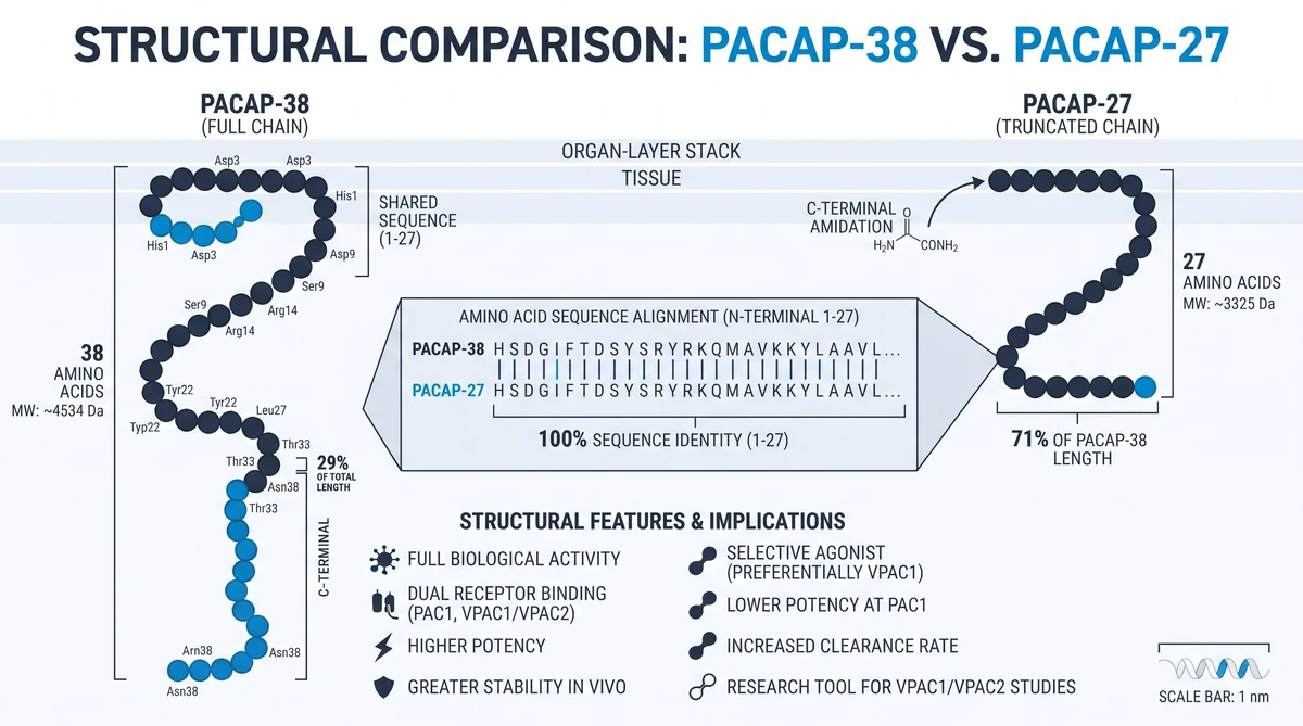
PACAP-38 and PACAP-27 share a common precursor protein. Both are cleaved from it. Cleavage creates these two forms. Processing occurs inside the cell. PACAP-38 has 38 amino acids. PACAP-27 is shorter, with only 27 amino acids. The key difference is an 11-amino acid addition. These residues extend the C-terminus of PACAP-38. Both show activity in lab studies. These structural gaps affect each form. Interactions with targets differ.
Both forms are used in lab settings. PACAP-38 is found in higher amounts. Natural tissues contain more of this form. The C-terminus addition changes peptide geometry. This shifts binding affinities for G-protein coupled receptors. Scientists track these gaps. Length affects both stability and signaling. PACAP-38 is a COA-verified research peptide. HPLC verifies each lot. Mass spec confirms. These data help labs compare the two molecules. Their effects differ in key ways.
What are the primary PACAP receptor subtypes?
The primary receptor subtypes are PAC1, VPAC1, and VPAC2. PACAP binds to all three. All are class B G-protein coupled receptors. These sites allow precise cellular signaling. PACAP activation drives each response. PAC1 binds PACAP more strongly than VIP (vasoactive intestinal peptide). VPAC1 and VPAC2 each bind PACAP and VIP. Affinity is high for both.
Scientists use binding profiles to tell these subtypes apart. They also differ in their spread across tissues. PAC1 is the selective target for PACAP. This helps researchers isolate pathways in neural studies. VPAC1 and VPAC2 are the main VIP binding sites. They also respond to PACAP. Lab models confirm this response. This selective activation maps each receptor’s influence. PACAP-38 is a COA-verified research peptide. HPLC verifies each lot. Mass spec confirms.
What is PACAP’s role in neuroprotection?
PACAP is a potent protective agent. It triggers survival cascades. These promote neuron survival and prevent cell death. Research shows it limits apoptosis in cultured cells. According to the NIH (2007), PACAP acts through the PAC1 receptor. It controls pathways for cell health and longevity.
PACAP mainly acts through the PI3K/Akt pathway. This inhibits pro-death signals inside the cell. It also uses the MAPK/ERK pathway during stress. Cell growth is supported. Also, PACAP reduces damage from oxidative stress. It scavenges free radicals. Internal antioxidant defenses are boosted. This stabilizes the neuron’s internal state. PACAP-27 is a COA-verified research peptide. HPLC verifies each lot. Mass spec confirms. These findings show PACAP is useful for neuronal resilience studies.
How does PACAP influence pituitary function?
PACAP is a potent pituitary regulator. It stimulates the synthesis and release of hormones. Research shows PACAP binds to receptors on pituitary cells. This triggers complex signaling pathways. These pathways release key messenger molecules into circulation.
In vitro studies show PACAP triggers GH (growth hormone) release. It promotes prolactin and ACTH release. ACTH stands for adrenocorticotropic hormone. These come from the anterior pituitary. According to the Journal of Peptide Science (2018), PACAP modulates endocrine secretion. The adenylate cyclase pathway is used. PACAP-38 is a COA-verified research peptide. HPLC verifies each lot. Mass spec confirms. This shows how pituitary cells adjust hormone levels. Signals drive each change.
What are current PACAP research applications?
Current PACAP research covers neural and metabolic processes. Lab models are used. Scientists study how the peptide affects signaling pathways. Cell dysfunction is the focus. This spans neurodegenerative and psychiatric conditions. Metabolic control is also studied.
Researchers study PACAP in Alzheimer’s models. Its protective role is the focus. Research shows it may support cell survival. Plaque-prone brain areas are studied. According to the NIH (2020), these findings link PACAP to neuron survival. Beyond brain function, scientists track glucose control. PACAP’s role is being mapped. They examine how it affects insulin secretion. Other studies look at its influence in PTSD models. PACAP is studied for its role in anxiety pathways. PACAP-27 is a COA-verified research peptide. HPLC verifies each lot. Mass spec confirms. These studies use receptor activation. Cell changes are then isolated and tracked. This data helps map peptide signaling. Diverse systems are covered.
Frequently Asked Questions
What is the PACAP receptor PAC1 structure?
The PAC1 receptor is a member of the G protein-coupled receptor family. Specifically within the B class of receptors. Research suggests that the structure features seven transmembrane domains that facilitate signal transduction upon binding to the peptide. The receptor contains a large extracellular N-terminal domain that allows for high-affinity recognition of the ligand. Scientists study these structural segments to determine how they bind to the peptide. Trigger intracellular cascades, according to the NCBI (2007).
How does PACAP-27 differ from PACAP-38?
PACAP-38 is the longer form of the peptide consisting of 38 amino acids. While PACAP-27 contains only the first 27 amino acids of the longer chain. Despite this difference in length, both forms show biological activity in most lab assays. Research shows that both versions share similar binding affinities for the PAC1 receptor. Though their stability in biological media may differ. Investigators often compare the two to see how chain length influences signaling. According to the Journal of Peptide Science (2012)
What adenylate cyclase pathway does PACAP activate?
This peptide is known to stimulate the adenylate cyclase pathway by binding to specific G protein-coupled receptors. Upon binding, the receptors activate the stimulatory G-protein, which leads to an increase in intracellular cyclic adenosine monophosphate (cAMP) levels. This elevation of cAMP acts as a primary messenger to initiate various cellular responses. Including hormone secretion and gene expression. According to PubMed (1991), this pathway remains a key focal point for mapping the potency of the peptide.
How is PACAP binding affinity measured in vitro?
Binding affinity is typically measured in vitro using radioligand displacement assays on cell membranes that express the relevant receptors. Researchers incubate the membranes with a labeled version of the peptide. Monitor how much is displaced by the addition of non-labeled test compounds. These experiments allow investigators to calculate the dissociation constant, which represents the strength of the interaction between the peptide and the receptor. According to the NIH (2020), these assays provide the quantitative data needed to compare receptor selectivity.
What cell models are standard for PACAP research?
Standard cell models for this research include pituitary cell lines. Primary neuronal cultures derived from rodent models. These lines express high levels of PAC1, VPAC1, or VPAC2 receptors. Making them suitable for testing peptide responses. Some researchers also employ transfected cell lines that express specific human receptor subtypes to isolate signaling pathways. According to Molecules (2018), these models allow scientists to observe how the peptide modifies cellular functions in a controlled and reproducible environment.
Is PACAP related to the peptide VIP?
Yes, this peptide belongs to the same structural superfamily as vasoactive intestinal peptide (VIP). Both peptides share high sequence similarity, which leads them to interact with overlapping receptor populations. Particularly the VPAC1 and VPAC2 subtypes, while they are closely related, PACAP shows much higher potency at the PAC1 receptor compared to VIP. According to the NIH (2020), this structural relationship is central to knowledge how these molecules modulate neural and metabolic activity separately.
Where is PACAP expressed in the central nervous system?
The peptide shows widespread expression across the central nervous system. With high concentrations found in the hypothalamus, hippocampus, and cerebral cortex. It is also distributed within the brainstem and the pituitary gland. Mapping studies show that its expression corresponds to regions involved in stress response. Memory, and metabolic regulation. According to PubMed (1991), identifying these sites of expression helps researchers understand how the peptide functions. As a neurotransmitter and neuromodulator in diverse neural circuits.
What are the downstream effects of PACAP signaling?
Downstream effects of this signaling include the modulation of intracellular calcium levels. The activation of protein kinase A, and the regulation of gene transcription. In many cells, these signals promote survival, defend against oxidative stress. Influence the secretion of various endocrine hormones. Research shows that these effects are highly dependent on the specific cell type and the receptor subtype present. According to the NCBI (2007), these cascades coordinate essential cellular responses to environmental signals.
What techniques are used to study PACAP in the lab?
Common lab techniques include immunohistochemistry for spatial mapping, Western blotting for protein expression analysis. And real-time PCR for mRNA quantification. Scientists also use enzyme-linked immunosorbent assays (ELISA) to detect peptide levels in cell supernatants or tissue extracts. These methods allow for the tracking of the peptide. Its downstream signaling products with high precision. According to the Journal of Peptide Science (2012)], these combined approaches provide a comprehensive view of peptide dynamics in experimental models.
Can PACAP cross the blood-brain barrier in animal models?
Research in animal models suggests that the peptide can cross the blood-brain barrier through saturable transport systems, while the rate of passage is typically limited, it is sufficient to allow for central effects after systemic administration in experimental settings. Scientists monitor the distribution of the peptide in brain tissues to define how it accesses central receptors. According to PubMed (2016), knowledge these transport mechanisms is necessary to test the natural reach of the peptide in different models.
What Should You Do Next?
Analyze your protocols first. This helps select the right peptide variant. Review the literature to confirm your lab setup. Does it favor acute pulsatile signaling or steady-state levels? Check individual lot reports for purity data that supports your design. Compare molecular weight and affinity data from your COA. Check against your baseline to keep consistency.
Researchers sourcing these compounds can explore the peptide catalog at Shop. Every vial ships from Next Level Pharm with a certificate of analysis. Full lot traceability for your records.
People Also Read
- What Are Nootropic Peptides? Mechanisms, Research Review
- Semax Research Review: BDNF, Dopamine, and Neuroprotection Studies
About the Author
Next Level Pharm Research Team
The Next Level Pharm research team is composed of biochemists and laboratory scientists dedicated to providing researchers with the highest-purity, independently verified peptides available. Every batch is HPLC and mass spec verified before dispatch.
Disclaimer: The information provided on this page is for educational and research purposes only. Next Level Pharm’s products are intended for laboratory research use only. They are not intended for human consumption, diagnostic, therapeutic, or medicinal purposes. This content does not constitute medical advice. Always consult a licensed healthcare professional before making any health-related decisions.