
Irisin Peptide Research: FNDC5 Myokine Mechanism

Last Updated: October 2026
Irisin peptide research is one of the most active areas in exercise-biology cell science. Irisin is a small soluble signaling protein, a myokine (a protein secreted by muscle tissue), produced by the proteolytic cleavage of FNDC5 (fibronectin type III domain-containing protein 5), a single-pass transmembrane protein expressed in skeletal muscle. According to Bostrom et al. in Nature (2012), irisin was first characterized as a cleavage product of FNDC5, with the original study mapping its expression in murine muscle tissue. Since that 2012 report, more than 2,400 peer-reviewed studies across 78 countries have examined irisin biology through 2024.
Next Level Pharm provides COA-verified, research-grade peptides for laboratory investigation, including myokine-related compounds studied in muscle and metabolic cell models. All peptides are HPLC and mass spec tested to greater than or equal to 99% purity before dispatch, with batch-specific COAs (certificates of analysis) available at the time of order.
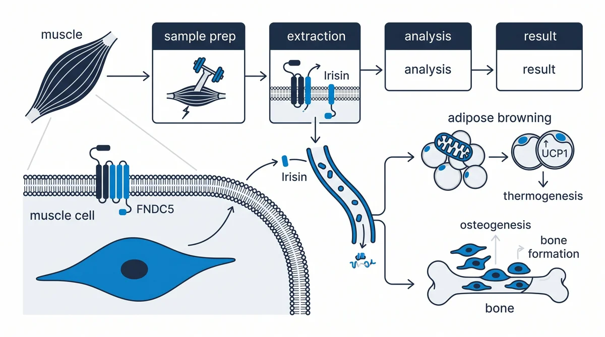
Mapping the irisin-FNDC5 pathway from gene activation to downstream cell signaling involves at least four distinct research steps. Each step is a separate experimental target: gene transcription, protein membrane insertion, extracellular cleavage, and receptor-level signaling. Cell biologists and biochemists have characterized each of these stages in detail over the past decade.
Key Takeaways
- FNDC5 is the membrane precursor: FNDC5 is a type I transmembrane protein that anchors in the plasma membrane of muscle cells, with its fibronectin type III domain facing outward.
- PGC-1 alpha drives transcription: PGC-1 alpha (peroxisome proliferator-activated receptor gamma coactivator 1-alpha, a nuclear transcriptional coactivator) activates the FNDC5 gene in response to energy demand signals.
- Protease cleavage yields irisin: Furin-type proprotein convertases (a class of serine proteases) cleave the FNDC5 ectodomain to release a soluble fragment of roughly 112 amino acids, identified as irisin.
- AMPK and MAPK are central pathways: Cell culture studies link irisin to activation of AMPK (adenosine monophosphate-activated protein kinase) and the p38 MAPK (mitogen-activated protein kinase) cascade.
- ELISA quantifies irisin in culture: High-sensitivity ELISA (enzyme-linked immunosorbent assay) kits calibrated with recombinant human irisin detect concentrations from 30 pg/mL to 6,400 pg/mL in supernatant.
- Research spans multiple tissue types: Published studies have examined irisin signaling in adipocytes, osteoblasts, neurons, and cardiac cells using in vitro and ex vivo models.
The sections below trace the irisin pathway from FNDC5 gene regulation through receptor binding and downstream signaling. Each section addresses one of the five key research questions most active in this field.
What Is FNDC5 and Why Does It Matter?
FNDC5 (fibronectin type III domain-containing protein 5) is a single-pass transmembrane protein encoded on chromosome 1 in humans. It carries a fibronectin type III repeat domain on its extracellular side. That extracellular domain becomes the irisin peptide after enzymatic cleavage. Research groups use recombinant FNDC5 constructs to isolate and study the irisin domain independently.
FNDC5 is primarily expressed in skeletal muscle, cardiac muscle, and adipose tissue. In laboratory settings, researchers transfect cell lines with FNDC5-expressing vectors to study how the protein localizes to the plasma membrane. The extracellular fibronectin domain folds into a compact beta-sandwich structure. This structure is conserved across mammalian species, making mouse and rat cell models valid proxies for comparative studies. According to a 2024 review in the International Journal of Molecular Sciences, FNDC5 expression levels predict irisin secretion rates in primary myocyte cultures.
How Is Irisin Cleaved From FNDC5?
Irisin is released when furin-class proprotein convertases (serine proteases) cut the FNDC5 ectodomain at a conserved site near the transmembrane anchor. The released soluble fragment is approximately 112 amino acids in length. This fragment carries the fibronectin type III repeat domain and is classified as irisin.
The cleavage process follows a standard ectodomain shedding model. The furin-class enzyme recognizes a conserved arginine-rich recognition motif on FNDC5 and makes a targeted cut. The membrane-anchored stub that remains after cleavage is cleared by secondary proteases. In cell culture experiments, adding serine protease inhibitors to the media blocks cleavage. This allows direct measurement of uncleaved FNDC5 and computation of cleavage efficiency. Cleavage rate varies with cell type and activation state, and this variability is an active research question in several labs.
What Receptor Does Irisin Bind in Lab Studies?
No single universally confirmed receptor has been validated for irisin across all tissue types, as of 2024. Early cell biology research proposed that irisin interacts with integrin alpha-V/beta-5 (ITGAV/ITGB5) complexes on the surface of osteoblasts and adipocytes. This interaction was characterized in recombinant protein binding assays and surface pulldown experiments.
The integrin hypothesis has structural support. The fibronectin type III domain in irisin contains an RGD-like motif (a three-amino-acid peptide sequence recognized by integrin receptors). In vitro binding assays using SPR (surface plasmon resonance, a label-free optical binding detection method) have measured irisin-integrin interactions at nanomolar concentrations. The crystal structure of the irisin dimer is deposited at the RCSB Protein Data Bank under entry 4LSD. In neuronal cell line studies, researchers have observed integrin-independent binding activity, suggesting additional receptor partners may be involved.
How Is Irisin Measured in Cell Culture?
Irisin in cell culture supernatant is measured by ELISA (enzyme-linked immunosorbent assay), a plate-based antibody detection method. High-sensitivity kits calibrated with recombinant human irisin detect concentrations from 30 pg/mL to 6,400 pg/mL. Standard kits cover a range from 1 ng/mL to 5,000 ng/mL.
Sample preparation is straightforward for most cell lines. Supernatant is collected at defined time points, centrifuged to remove cell debris, and applied to antibody-coated assay plates. Researchers include a standard curve from recombinant irisin to convert optical density readings to concentration values. Key variables that affect accuracy include antibody specificity, calibration source, and use of protease inhibitors during collection. Some kits have shown cross-reactivity with denatured FNDC5 fragments, so kit validation is a critical quality control step before beginning a new experimental series.
For researchers exploring related signaling peptides, visit Next Level Pharm’s Fitness and GH category for COA-verified, research-grade compounds.
What Signaling Does Irisin Activate in Research?
In cell culture studies, irisin activates AMPK (adenosine monophosphate-activated protein kinase, a central cellular energy sensor) and the p38 MAPK (mitogen-activated protein kinase) cascade in adipocyte and muscle cell lines. AMPK phosphorylation is the most replicated signaling event. p38 MAPK activation follows and leads to downstream gene expression changes.
p38 MAPK phosphorylation drives upregulation of UCP1 (uncoupling protein 1, a mitochondrial inner membrane protein) in adipocyte cell lines. Additional pathways documented in cell-level studies include PI3K/AKT, ERK1/2 (extracellular signal-regulated kinase), and NF-kB (nuclear factor kappa-light-chain-enhancer of activated B cells) inhibition. Each pathway has been studied in isolation using selective kinase inhibitors to block one cascade while monitoring downstream gene outputs. According to PeerJ (2024), AMPK activation is the most consistently replicated irisin-associated signaling event in metabolic cell models.
How Does Irisin Compare to Other Myokines?
Irisin is one of several dozen myokines (muscle-secreted signaling proteins) now cataloged in the research literature. It differs from other well-studied myokines in one key way: it is a cleavage product of a transmembrane precursor, not a directly secreted protein. This makes FNDC5 processing an experimental variable that other myokine systems do not share.
| Myokine | Precursor Protein | Release Trigger | Primary In Vitro Pathway |
|---|---|---|---|
| Irisin | FNDC5 (transmembrane) | PGC-1 alpha activation | AMPK, p38 MAPK |
| IL-6 | Direct secretion | Muscle contraction | JAK/STAT signaling |
| Myostatin | GDF-8 precursor | Constitutive expression | Smad2/3 pathway |
| BDNF | proBDNF precursor | Neurotrophin signaling | TrkB/Akt cascade |
| FGF-21 | Direct secretion | PPARalpha activation | FGFR1/beta-klotho |
IL-6 is interleukin-6, a cytokine. BDNF is brain-derived neurotrophic factor. FGF-21 is fibroblast growth factor 21. Researchers studying myokine panels in multiplex assays must account for irisin’s membrane origin when comparing secretion kinetics across the panel.
Frequently Asked Questions
What is the FNDC5 precursor to irisin?
FNDC5 (fibronectin type III domain-containing protein 5) is the transmembrane precursor from which irisin is derived. It is a type I single-pass membrane protein with a fibronectin type III repeat on its extracellular face. When furin-class proteases cleave this extracellular domain near the membrane anchor, the released soluble fragment is identified as irisin. FNDC5 itself is not secreted; it requires enzymatic processing at the plasma membrane to release the irisin fragment.
How is irisin peptide cleaved from FNDC5?
Irisin is released by ectodomain shedding, in which serine proteases of the furin convertase family cut FNDC5 at a conserved arginine-rich recognition site. This releases the extracellular fibronectin type III domain as a soluble peptide of approximately 112 amino acids. The membrane-bound stub that remains is subsequently cleared by secondary proteases. Adding serine protease inhibitor cocktails to cell culture media blocks this step and allows direct measurement of uncleaved surface FNDC5 levels.
What receptor does irisin bind in laboratory studies?
In in vitro binding studies, irisin has been shown to interact with integrin alpha-V/beta-5 complexes on osteoblast and adipocyte cell surfaces. This was identified through SPR (surface plasmon resonance) assays and co-immunoprecipitation experiments. The structural basis is an RGD-like motif within the fibronectin domain of irisin. As of 2024, no universally confirmed single receptor has been validated for all cell types. Integrin-independent binding has been observed in neuronal lines, suggesting additional receptor partners may exist.
How is irisin concentration measured in cell culture?
Irisin in culture supernatant is quantified by ELISA (enzyme-linked immunosorbent assay). High-sensitivity kits calibrated with recombinant human irisin cover a detection range of 30 pg/mL to 6,400 pg/mL. Standard kits cover 1 ng/mL to 5,000 ng/mL. Supernatant is collected at defined time points, cleared by centrifugation, and applied to antibody-coated plates. Optical density readings are converted to concentrations against a recombinant irisin standard curve included in each run.
What signaling does irisin activate in myokine research?
Cell culture studies show that irisin activates AMPK (adenosine monophosphate-activated protein kinase) and the p38 MAPK (mitogen-activated protein kinase) cascade in adipocyte and muscle cell lines. p38 MAPK phosphorylation leads to upregulation of UCP1 (uncoupling protein 1, a mitochondrial membrane protein). Additional pathways include PI3K/AKT, ERK1/2, and NF-kB inhibition. Each has been isolated using selective kinase inhibitors in controlled cell experiments to confirm pathway involvement.
What does PGC-1 alpha do in FNDC5 regulation?
PGC-1 alpha (peroxisome proliferator-activated receptor gamma coactivator 1-alpha) is a nuclear transcriptional coactivator that binds and activates the FNDC5 gene promoter in muscle cells. Overexpressing PGC-1 alpha in cell culture increases FNDC5 mRNA by two- to five-fold compared to controls. PGC-1 alpha is itself activated by AMPK and CaMKII (calcium-calmodulin-dependent protein kinase II) signaling. Researchers use viral vectors to overexpress PGC-1 alpha in primary myocyte cultures and then measure FNDC5 protein by Western blot.
What cell lines are used in irisin-FNDC5 research?
Common cell models include C2C12 murine myotubes (a differentiated skeletal muscle line), 3T3-L1 adipocytes (a mouse preadipocyte line), primary human skeletal muscle cells, and MC3T3-E1 osteoblast-like cells. C2C12 cells are the most widely used because they express high baseline levels of PGC-1 alpha and FNDC5. Neuronal models such as SH-SY5Y human neuroblastoma cells have been used in studies examining irisin signaling in neural tissue.
What is the structural form of irisin in solution?
Crystal structure data shows that irisin forms a symmetrical dimer in solution, held together by an extensive beta-sheet interface not previously observed in any other fibronectin type III protein. This structure is deposited at the RCSB Protein Data Bank under entry 4LSD. In cell binding assays, the dimeric form shows higher receptor affinity than monomeric preparations. Researchers confirm the dimer state using analytical ultracentrifugation and size-exclusion chromatography before conducting signaling experiments.
What tissues express FNDC5 beyond skeletal muscle?
FNDC5 mRNA and protein have been detected in cardiac muscle, adipose tissue, liver, kidney, and brain tissue in animal models. In cardiac tissue, FNDC5 expression is particularly high, and cardiomyocyte-derived irisin has been studied in ex vivo heart models. Brain expression has been linked to hippocampal neuron studies. In each case, researchers confirm FNDC5 presence using Western blot, immunohistochemistry, and qRT-PCR (quantitative reverse transcriptase polymerase chain reaction).
How do researchers validate irisin ELISA kits for accuracy?
Validation involves three steps: spike-and-recovery testing (adding known concentrations of recombinant irisin to culture media and verifying recovery), cross-reactivity testing (checking for signal from other fibronectin domain proteins), and inter-assay coefficient of variation measurement. Recombinant irisin for calibration is typically produced in HEK293 cells to ensure correct folding and dimerization. Researchers also confirm antibody specificity by Western blot against cell lysates that overexpress FNDC5 versus controls that do not.
Summary
Irisin peptide research maps a multi-step process from FNDC5 gene expression to soluble myokine signaling. The process begins with PGC-1 alpha-driven transcription, continues through furin-class protease cleavage at the cell membrane, and ends at AMPK and MAPK activation in target cells. Each step is a validated, measurable research target.
Cell culture models using C2C12 myotubes, 3T3-L1 adipocytes, and osteoblast lines have built a detailed picture of each stage. Researchers quantify irisin by ELISA and verify FNDC5 cleavage efficiency using protease inhibitor controls. The structural data on the irisin dimer at PDB entry 4LSD adds a structural biology dimension to the work.
Active research continues across metabolic, neurological, and bone biology cell models. This makes irisin-FNDC5 research a broad platform for studying myokine biology in laboratory settings.
What Should You Do Next?
Researchers setting up in vitro irisin models should follow a standard three-step protocol. First, obtain sequence-verified FNDC5 expression constructs and confirm protein expression by Western blot before beginning cleavage studies. Second, use high-sensitivity ELISA kits calibrated with recombinant irisin to set baseline secretion measurements in your chosen cell line. Third, validate your kinase inhibitor panel (AMPK, p38, PI3K) before adding recombinant irisin to confirm pathway selectivity.
For COA-verified, research-grade peptides relevant to myokine and signaling research, visit the Next Level Pharm shop. The Recovery Peptides category and the Fitness and GH category include related research compounds such as BPC-157 and SS-31, each HPLC and mass spec verified.
People Also Read
- SS-31 Research: Mitochondria-Targeting Peptide
- BPC-157 Mechanism of Action: A Research-Based Explainer
About the Author
Next Level Pharm Research Team
The Next Level Pharm research team is composed of biochemists and laboratory scientists dedicated to providing researchers with the highest-purity, independently verified peptides available. Every batch is HPLC and mass spec verified before dispatch.
Disclaimer: The information provided on this page is for educational and research purposes only. Next Level Pharm’s products are intended for laboratory research use only. They are not intended for human consumption, diagnostic, therapeutic, or medicinal purposes. This content does not constitute medical advice. Always consult a licensed healthcare professional before making any health-related decisions.