
Neurotensin Peptide Research: NTS1 Receptor Studies

Last Updated: October 2026
Neurotensin peptide research is an active area of GPCR biology that examines how a 13-amino acid neuropeptide activates the NTS1 receptor (neurotensin receptor type 1) to trigger intracellular signaling cascades. First isolated from bovine hypothalamic tissue in 1973, neurotensin is expressed in both the central nervous system (CNS) and peripheral tissues. Published in vitro data show that full-length neurotensin binds NTS1 in the low nanomolar range, making it a reliable pharmacological tool for receptor binding and signaling studies.
Next Level Pharm supplies research-grade neurotensin peptide for laboratory applications. Each batch carries a certificate of analysis (COA), confirming purity by HPLC (high-performance liquid chromatography) and mass spectrometry. Lyophilized vials ship with full lot documentation, supporting traceable in vitro studies from binding assays through signaling pathway analysis.
Neurotensin receptor studies span several model systems. Researchers work with recombinant cell lines, primary neuronal cultures, and lipid nanodisc preparations. Each system answers different questions about receptor structure, G protein coupling, and ligand pharmacology. The Key Takeaways below connect the main findings before the detailed sections.
Key Takeaways
- NTS1 Structure: NTS1 is a class A GPCR with a seven-transmembrane domain fold. The orthosteric binding pocket is formed by residues in transmembrane helices 3, 5, 6, and 7.
- Binding Fragment: Neurotensin engages NTS1 primarily through its C-terminal hexapeptide (residues 8 to 13, called NT8-13). This fragment retains full receptor affinity in competitive binding assays.
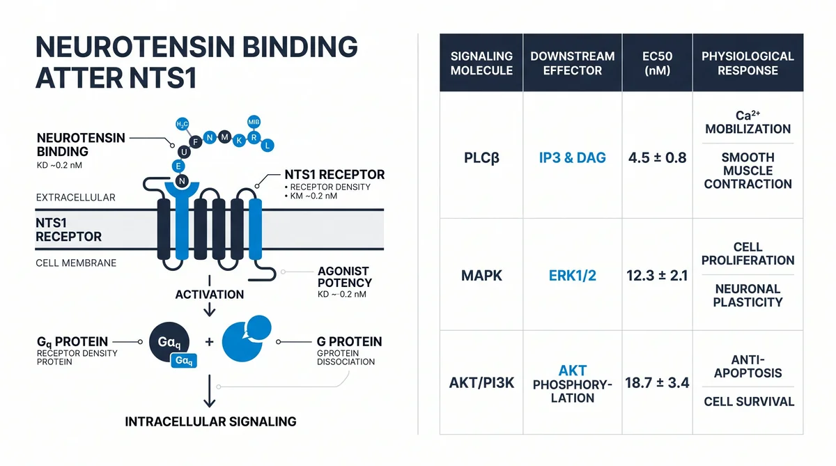
- G Protein Coupling: NTS1 couples to Gq and Go protein families. Gq activation leads to phospholipase C-beta (PLC-beta) stimulation, IP3 production, and intracellular calcium release.
- Beta-Arrestin Pathway: Beta-arrestin-1 is recruited to NTS1 after G protein activation. This drives receptor internalization through clathrin-coated pits and activates MAP kinase (mitogen-activated protein kinase) pathways.
- In Vitro Models: CHO-K1 and HEK293 cells stably expressing NTS1 are standard research hosts. BRET (bioluminescence resonance energy transfer) assays quantify G protein and beta-arrestin coupling in live cells.
- Purity Requirements: Radioligand binding assays require peptide purity above 95%. Research-grade neurotensin at 99%+ purity reduces assay noise and supports reproducible displacement data.
These six points frame the detailed sections below. Each section cites published research for readers who want primary source data.
What Is the NTS1 Neurotensin Receptor Structure?
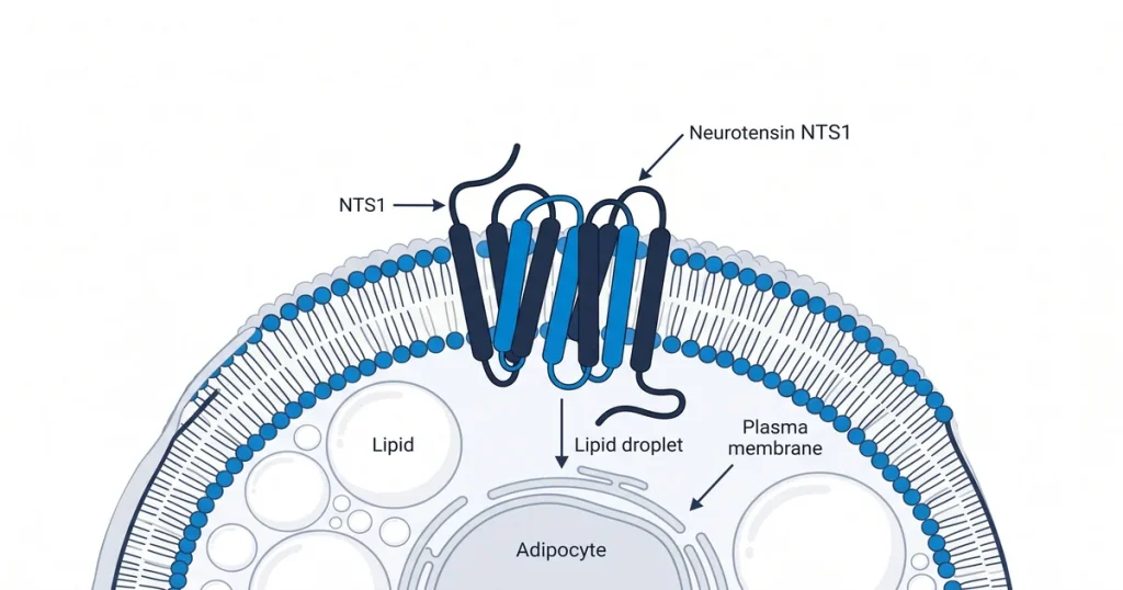
NTS1 belongs to the class A GPCR superfamily. Seven alpha-helical transmembrane (TM) segments form the receptor core, connected by intracellular loops (ICLs) and extracellular loops (ECLs). The orthosteric binding pocket sits within the extracellular half of the TM bundle. Extracellular loop 2 (ECL2) folds over the pocket and contributes key contacts for ligand docking.
According to White et al. via PubMed Central (2023), cryo-EM structures of NTS1 in complex with Gq and Go protein heterotrimers reveal an extended network of protein-protein contacts at the receptor-transducer interface. Structures resolved in lipid bilayers show greater detail than those in detergent micelles, underscoring the role of membrane environment in receptor conformation. This structural data guides the design of NTS1-targeted research probes and explains how different transducers engage the same receptor core.
How Does Neurotensin Interact With the NTS1 Receptor?
Neurotensin docks to NTS1 through its C-terminal hexapeptide fragment, NT8-13. This fragment contains tyrosine, leucine, and arginine residues that make direct contacts with binding pocket residues in TM helices 3, 5, 6, and 7. Full-length neurotensin and NT8-13 show comparable binding affinities in competitive displacement assays, making NT8-13 a preferred research tool due to its smaller size and easier synthesis.
The binding process starts with an initial extracellular entry conformation of the receptor. Conformational tightening follows as the ligand settles into the orthosteric pocket. Research using BRET-based assays in CHO-K1 cells confirms that neurotensin binding leads to rapid Gq protein coupling. Beta-arrestin-1 engagement follows G protein activation and drives receptor endocytosis through clathrin-coated pits. Internalization is temperature-dependent and blocked by endocytosis inhibitors, confirming it occurs through active vesicle trafficking.
What Signaling Pathways Does NTS1 Activation Trigger?
NTS1 activation routes primarily through Gq proteins. Gq activates phospholipase C-beta (PLC-beta), which cleaves PIP2 (phosphatidylinositol 4,5-bisphosphate) into two second messengers: IP3 (inositol trisphosphate) and DAG (diacylglycerol). IP3 triggers calcium release from the endoplasmic reticulum. DAG activates PKC (protein kinase C), which phosphorylates multiple downstream targets.
Key signaling outputs measured in NTS1 in vitro studies:
- Calcium mobilization: Measured by FLIPR (fluorometric imaging plate reader) assay in HEK293 and CHO-K1 cells using calcium-sensitive dyes
- ERK1/2 phosphorylation: Extracellular signal-regulated kinase 1 and 2 activation through both G protein and beta-arrestin pathways, measured by Western blot
- Receptor internalization: Quantified by cell-surface ELISA on extracellular epitope antibodies or by confocal microscopy using fluorescently tagged NTS1
According to Krumm et al. in Biochemistry (2023), allosteric modulators of NTS1 can shift signaling toward beta-arrestin pathways while reducing G protein responses. This selective activation, called biased agonism, is an active focus of GPCR pharmacology. The study used cryo-EM structures of NTS1 ternary complexes with Gq and GoA to map allosteric sites at high resolution.
Planning NTS1 studies for your lab? Shop research-grade neuropeptides at Next Level Pharm for HPLC-verified, lyophilized peptides with full COA documentation and lot-level traceability.
How Is Neurotensin Receptor Binding Measured In Vitro?
Radioligand competition assays are the primary method for NTS1 binding affinity data. [125I]-labeled neurotensin or [3H]-NT serves as the tracer. Unlabeled test compounds displace the tracer to give Ki values (inhibition constants) that rank potency. Cell membrane preparations from NTS1-expressing cells provide the receptor source for assay plates.
Scintillation proximity assays (SPA) enable high-throughput binding in 96-well or 384-well format, reducing radioactive handling volume. Functional assays, such as the HTRF (homogeneous time-resolved fluorescence) IP-One assay, measure IP1 (inositol monophosphate) accumulation as a surrogate for Gq activation. BRET assays track beta-arrestin-2 recruitment in live cells without fixation or lysis. Each method provides a different view of receptor engagement: direct binding, G protein activation, or arrestin coupling.
According to researchers at Friedrich-Alexander University (2024), a comparative study of neurotensin(8-13) analogs at NTS1 and NTS2 receptors combined competitive displacement assays on purified membranes with BRET-based cellular assays. This combined approach provides both affinity and functional coupling data, supporting structure-activity relationship (SAR) programs for NT8-13 analogs.
What Cell Models Express NTS1 for Research Studies?
Several cell lines serve as standard NTS1 research hosts:
- HEK293 cells: Human embryonic kidney cells with low endogenous GPCR background. Transient or stable NTS1 transfection is straightforward. BRET and HTRF assays perform reliably in this background.
- CHO-K1 cells: Chinese hamster ovary cells, a widely used host for stably transfected GPCR lines. Stable CHO-K1-NTS1 lines show consistent neurotensin response in calcium assays.
- HCT116 cells: A colorectal cancer cell line that expresses NTS1 endogenously. Useful for studying receptor signaling in a native context without overexpression artifacts.
- Lipid nanodiscs: Membrane-mimetic preparations of purified NTS1 protein for NMR and cryo-EM studies where cellular context is not required.
| Cell Model | NTS1 Source | Primary Readout | Notes |
|---|---|---|---|
| HEK293 | Transfected | BRET, HTRF, calcium | Low endogenous GPCR background |
| CHO-K1 | Stable line | Calcium, IP1 | Reliable GPCR host cell |
| HCT116 | Endogenous | ERK, calcium | Native receptor context |
| Xenopus oocyte | mRNA injection | Electrophysiology | Ion channel coupling studies |
| Lipid nanodiscs | Purified protein | NMR, cryo-EM | Structural studies only |
How Do Lipids Influence NTS1 Receptor Dynamics?
Membrane lipid composition shapes NTS1 behavior beyond the receptor-ligand interaction. NMR spectroscopy studies show that PIP2 binding to the intracellular receptor surface adjusts the timescale of conformational exchange at the orthosteric pocket. This allosteric effect occurs without dramatically changing the overall receptor structural ensemble.
According to Uchanski et al. in Nature Communications (2023), the conformational dynamics of NTS1 differ from its static crystal structure. The receptor samples multiple states in the membrane, and lipid composition shifts which states are preferred. Cholesterol removal using methyl-beta-cyclodextrin disrupts receptor clustering and reduces agonist-stimulated calcium responses. Researchers account for this variable when comparing data across cell-based and nanodisc-based model systems.
Next Level Pharm provides COA-verified, HPLC-tested neuropeptides for brain peptide research. Researchers focused on tissue repair applications can also explore recovery peptide tools in the catalog.
Frequently Asked Questions
What is the molecular weight of neurotensin?
Neurotensin is a 13-amino acid peptide with a molecular weight of approximately 1,673 daltons. Its full sequence runs from pyroglutamate at position 1 through leucine at position 13. The C-terminal fragment NT8-13 has a molecular weight near 832 daltons and retains full NTS1 binding activity. Both the full-length peptide and NT8-13 are available as research-grade peptides for receptor binding and functional assays.
How many neurotensin receptor subtypes exist?
Three neurotensin receptor subtypes have been identified in mammals: NTS1, NTS2, and NTS3. NTS1 and NTS2 are both GPCRs with distinct affinity profiles and tissue distributions. NTS1 shows high affinity for neurotensin, with Kd values in the low nanomolar range. NTS3, also called sortilin, is a single-pass transmembrane protein from a separate receptor family. Most in vitro pharmacology work focuses on NTS1 and NTS2, which share structural similarity and partial ligand overlap.
What is biased agonism at NTS1?
Biased agonism refers to a ligand’s ability to favor one downstream signaling pathway over another at the same receptor. At NTS1, biased agonists can preferentially activate G protein coupling or beta-arrestin recruitment. Beta-arrestin biased compounds show different calcium mobilization profiles compared to G protein-biased tools in the same cell system. Understanding NTS1 signaling bias is an active area of receptor pharmacology, with cryo-EM studies in 2023 mapping allosteric sites that control pathway selection.
What is the role of neurotensin in the central nervous system?
In the CNS, neurotensin modulates dopamine signaling in key circuits. NTS1 receptors colocalize with dopamine neurons in the VTA (ventral tegmental area) and substantia nigra. Neurotensin released at these synapses adjusts dopamine neuron firing rates and dopamine release in the nucleus accumbens. These interactions make NTS1 a mechanistic research target for dopamine circuit studies. Research-grade peptide tools support in vitro dissection of these receptor-mediated effects.
Can neurotensin peptides be applied in animal research models?
Neurotensin peptides have been applied in rodent pharmacology studies through systemic administration and intracerebroventricular (ICV) infusion routes. These studies examine behavioral endpoints including locomotor activity, thermal response, and nociception using standard research protocols. All animal work requires proper institutional approval. Next Level Pharm’s lyophilized neurotensin peptide is intended for laboratory research use only and is not intended for human application.
What does NTS1 receptor binding affinity data look like?
NTS1 receptor binding affinity is expressed as Ki (inhibition constant) or Kd (dissociation constant) values. Full-length neurotensin shows Ki values of roughly 0.1 to 1.0 nM at recombinant human NTS1. NT8-13 analog affinity varies with modification, with some analogs showing Ki values below 0.5 nM. These data come from radioligand competition assays using [125I]-NT tracer on cell membrane preparations, analyzed by non-linear regression curve fitting.
What does COA verification include for research peptides?
COA (certificate of analysis) verification for research peptides includes HPLC purity analysis and mass spectrometry confirmation. HPLC separates the peptide from synthesis impurities on a reverse-phase column and reports purity as a percent of total peak area. Mass spectrometry confirms the measured molecular weight matches the theoretical value within accepted tolerances. Each COA carries a lot number that links the vial to its batch production record, supporting experimental traceability.
What is the difference between NTS1 and NTS2 in binding studies?
NTS1 and NTS2 share similar binding pocket architecture but differ in ligand affinity profiles. NTS1 binds the NT8-13 fragment with sub-nanomolar to low nanomolar affinity. NTS2 shows moderate affinity for NT8-13 and higher affinity for some synthetic peptide analogs. Receptor-selective compounds such as the NTS1 antagonist SR48692 allow researchers to attribute signaling responses to a specific subtype in cells expressing both receptors. Selectivity profiling is standard practice in neurotensin analog research programs.
How is NTS1 receptor internalization studied?
NTS1 internalization after agonist exposure is tracked by several methods. Confocal microscopy using fluorescently tagged NTS1 shows receptor redistribution from the plasma membrane to endosomal compartments. Cell-surface ELISA with antibodies against an extracellular epitope measures total surface receptor loss after agonist treatment. Beta-arrestin BRET assays provide indirect tracking of internalization in live cells. Internalization is temperature-dependent and blocked by endocytosis inhibitors, confirming active vesicle trafficking.
Where is NTS1 expressed in peripheral tissues?
NTS1 is expressed in several peripheral tissues including the gastrointestinal (GI) tract, adrenal glands, and certain cancer cell lines. Colorectal cancer cell lines such as HCT116 express functional NTS1 and respond to exogenous neurotensin with calcium mobilization and ERK phosphorylation. Pancreatic cancer lines have also been reported as NTS1-positive in vitro models. Peripheral expression patterns make NTS1 a subject of in vitro cancer biology research alongside its primary roles in neuroscience.
Summary
Neurotensin is a 13-amino acid neuropeptide that binds the NTS1 receptor, a class A GPCR, with low nanomolar affinity. Its C-terminal hexapeptide NT8-13 retains full binding activity and is widely used in assay formats. NTS1 activation drives Gq-coupled signaling through PLC-beta, IP3-mediated calcium release, and PKC stimulation. Beta-arrestin recruitment follows and drives receptor internalization through endocytic pathways. Common in vitro research models include HEK293, CHO-K1, and HCT116 cells. Structural studies using cryo-EM and NMR have resolved receptor conformational states and lipid allosteric effects in high-resolution detail.
What Should You Do Next?
Researchers starting NTS1 studies should confirm peptide purity before any assay. HPLC-verified, mass spec confirmed neurotensin peptides reduce variability in radioligand displacement and functional assays. Stable CHO-K1-NTS1 or HEK293-NTS1 cell lines provide consistent results for G protein and beta-arrestin coupling measurements. For structural work, lipid nanodisc preparations require purified NTS1 protein, and cryo-EM studies benefit from bilayer-embedded receptor rather than detergent-solubilized preparations.
Research steps to consider:
1. Select a cell model: HEK293 for low GPCR background; CHO-K1 stable line for reproducible calcium data; HCT116 for native receptor context.
2. Confirm neurotensin peptide purity by HPLC before running radioligand or functional assays.
3. Validate the primary readout (Ki from binding, EC50 from calcium) with one secondary assay (beta-arrestin BRET or ERK phosphorylation by Western blot).
People Also Read
- What is Selank? GABA Pathways, Mechanisms, and Study Findings
- Semax Research Review: BDNF, Dopamine, and Neuroprotection Studies
About the Author
Next Level Pharm Research Team
The Next Level Pharm research team is composed of biochemists and laboratory scientists dedicated to providing researchers with the highest-purity, independently verified peptides available. Every batch is HPLC and mass spec verified before dispatch.
Disclaimer: The information provided on this page is for educational and research purposes only. Next Level Pharm’s products are intended for laboratory research use only. They are not intended for human consumption, diagnostic, therapeutic, or medicinal purposes. This content does not constitute medical advice. Always consult a licensed healthcare professional before making any health-related decisions.