
GHK-Cu Skin Research: Before and After Study Data

Last updated: July 2026
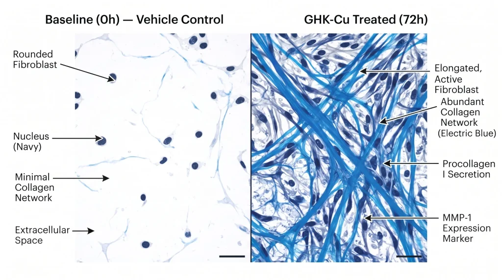
A GHK-Cu before and after study is a cell or animal model experiment. It compares skin gene markers before and after adding GHK-Cu (glycyl-L-histidyl-L-lysine copper(II), a copper-binding tripeptide). According to Pickart L et al. (2015) (PMID 25893651), GHK-Cu stimulates collagen and fibronectin gene expression in fibroblast cell models. All cited data come from cell and animal studies. No human outcomes are claimed or implied.
Next Level Pharm is a US-based supplier of research-grade GHK-Cu. Every lot is tested to ≥99% purity by HPLC and mass spec. A COA with lot number ships with each batch.
The term “before and after” in cell studies means measuring gene or protein markers at two time points. The first is baseline (before the compound). The second is after a set incubation period.
Key Takeaways
- Collagen Gene Markers Are Tracked: Collagen I and III gene expression levels are measured before and after GHK-Cu treatment in fibroblast cell models. Increased mRNA levels indicate upregulation of these genes.
- Fibronectin Is Also Measured: Fibronectin is a matrix glycoprotein. Cell model studies track fibronectin production after GHK-Cu treatment. Changes are measured by ELISA and Western blot.
- Keratinocyte Migration Is Studied: Scratch assays in keratinocyte cultures measure how quickly cells close a gap. GHK-Cu is added to treated wells. Migration speed is compared to vehicle control.
- Hair Follicle Cell Models Exist: In vitro hair follicle studies treat dermal papilla cells with GHK-Cu and measure gene markers for hair follicle growth phases. These are in vitro findings only.
- No Human Outcomes Are Proven: All GHK-Cu skin data cited here come from cell and animal models. No clinical trials have confirmed these effects in humans. The compound is for lab research use only.
Understanding which markers are measured and how the study is set up helps researchers evaluate the available literature and design appropriate protocols.
What Skin Markers Are Studied with GHK-Cu?
GHK-Cu cell studies track multiple gene and protein markers. The most common are collagen type I, collagen type III, and fibronectin. These are extracellular matrix (ECM) proteins produced by fibroblasts in skin tissue.
Other markers include matrix metalloproteinase (MMP) enzymes. MMPs break down ECM components. Some GHK-Cu studies track MMP-1 and MMP-2 levels. Lower MMP activity after treatment means less ECM breakdown in the model.
According to Pickart L (2008) (PMID 18561291), GHK-Cu changed collagen and fibronectin gene expression in human fibroblast cell cultures. These findings are from in vitro models only and cannot be extrapolated to human skin outcomes.
What Collagen Changes Appear in Cell Models?
Collagen is the main structural protein in skin tissue. In fibroblast cell models, collagen I and III are the two main types tracked. Studies use RT-PCR (reverse transcription polymerase chain reaction) to measure mRNA levels of these collagen genes.
In GHK-Cu studies, treated fibroblasts show increased collagen I and III mRNA compared to vehicle control in some in vitro models. The increase is measured as a fold change from baseline (before treatment). A fold change of 1.5 to 2.0 means collagen gene expression increased 50 to 100 percent above baseline in that model.
These are gene expression findings in cell culture. They do not equal tissue-level collagen production in living organisms.
How Do Fibroblasts Respond to GHK-Cu?
Fibroblasts are the main collagen-producing cells in connective tissue. In cell culture, fibroblasts are seeded in a dish and allowed to attach. GHK-Cu is added at a known concentration. After a set incubation time (typically 24 to 72 hours), RNA and protein are collected for analysis.
Researchers measure mRNA by RT-PCR and protein by ELISA or Western blot. Both methods compare treated cells to untreated controls. Changes are expressed as a ratio or fold change. Statistical significance is required for the result to be reported in peer-reviewed research.
GHK-Cu is available as a COA-verified research compound. Every lot is tested by HPLC and mass spec.
What Hair Follicle Data Exist?
Dermal papilla cells are a type of specialized skin cell in the hair follicle base. In cell models, dermal papilla cells are grown in culture and treated with GHK-Cu at various concentrations. Gene markers for follicle growth phases are measured by RT-PCR and protein assay.
According to Pickart L et al. (2018) (doi:10.3390/biomedicines6020039), GHK-Cu is studied in cell and animal models for skin structure gene marker changes. Hair follicle dermal papilla data are in vitro only. No clinical hair outcome data are available.
Some animal model studies also use topical application of GHK-Cu to rodent skin and measure histological markers of hair follicle activity. These are animal model findings only.
Shop research peptides. GHK-Cu is available with full COA documentation on every batch.
What Wound Model Data Exist?
In wound model studies, a scratch or circular wound is made in a cell monolayer (or in a rodent skin model). Measurements are taken at baseline (before treatment) and at set time points after GHK-Cu is added to treated groups.
Cell wound assays use keratinocytes or fibroblasts. Wound closure rate is measured by imaging at 12, 24, and 48 hours. Treated cells are compared to vehicle control cells. The rate of wound closure in treated vs. control groups is the main output.
Animal wound models use rodents with standardized skin wounds. Histological sections are taken at several time points after compound application. Collagen fiber density, cell density, and gene marker levels are measured in tissue sections. These are animal model findings only.
How Long Do Study Timelines Run?
Cell model studies typically run for 24 to 72 hours. This is enough time for gene expression changes to be measured by RT-PCR. Protein-level changes (by ELISA or Western blot) may need 48 to 96 hours.
Animal model studies run for longer timelines. Wound model studies in rodents may run for 7 to 21 days. Tissue is collected at several time points. Histological and gene marker data are compared across time points.
A “before and after” design always requires a baseline measurement and at least one post-treatment measurement. The interval between measurements depends on the marker being tracked.
Frequently Asked Questions
What Does GHK-Cu Stand For?
GHK-Cu stands for glycyl-L-histidyl-L-lysine copper(II). It is a copper-binding tripeptide made up of three amino acids: glycine (G), histidine (H), and lysine (K). The copper ion (Cu) binds to this tripeptide. It is found naturally in human plasma, saliva, and urine. GHK-Cu is studied in cell and animal models for its effects on collagen, fibronectin, and keratinocyte gene markers. All research data cited here come from in vitro and animal studies only. The compound is for lab research use only.
What Is Collagen Type I and III?
Collagen type I is the most abundant collagen in skin and connective tissue. It forms strong fibers that give skin its structural strength. Collagen type III is a thinner fiber found alongside type I. It contributes to skin elasticity in younger tissue. In fibroblast cell studies, both types are measured by RT-PCR as mRNA levels. An increase in collagen I and III mRNA means the collagen genes are more active in the cell model.
What Is Fibronectin?
Fibronectin is a large glycoprotein in the extracellular matrix (ECM). It helps cells attach to the matrix and move through tissue. In skin cell models, fibronectin production is measured by ELISA or Western blot. GHK-Cu studies track fibronectin levels in treated vs. control fibroblast cultures. Higher fibronectin levels indicate more ECM protein production in the cell model. This is an in vitro finding only.
What Is a Scratch Assay?
A scratch assay makes a straight gap in a monolayer of cells in a culture dish. The gap is photographed at time zero (before treatment). GHK-Cu is then added to treated wells. The gap is photographed again at 12, 24, and 48 hours. The rate at which cells close the gap is measured by image analysis software. This rate is compared between treated and control groups. The scratch assay is a standard in vitro method for studying cell migration.
What Is RT-PCR?
RT-PCR (reverse transcription polymerase chain reaction) is a lab method for measuring gene expression. It converts mRNA into cDNA (complementary DNA) and then amplifies a specific gene sequence. The amount of amplified product is proportional to the amount of mRNA in the original sample. In GHK-Cu studies, RT-PCR measures how much collagen I, collagen III, or other gene mRNA is present in treated vs. control cells.
What Concentration Is Used in Cell Studies?
GHK-Cu concentration in cell studies varies by protocol. Common ranges are 1 nanomolar to 10 micromolar. Studies use multiple concentrations to find a concentration-response curve. The concentration that produces the largest change with minimal cell toxicity is reported as the effective concentration. Researchers use cell viability assays (MTT or CCK-8) alongside gene marker assays to confirm concentrations are not toxic to the cell model.
Can GHK-Cu Cell Data Predict Human Skin Results?
No. Cell model data cannot directly predict human skin outcomes. The cell culture environment is much simpler than living skin tissue. Gene expression changes in a cell culture dish do not equal tissue-level changes in a human body. Many compounds that show effects in cell models fail to show the same effects in animal models or clinical trials. All GHK-Cu data cited here come from cell and animal models only. No human outcome claims are made.
What Is an ELISA?
ELISA (enzyme-linked immunosorbent assay) is a plate-based method for measuring the amount of a specific protein in a sample. A plate is coated with an antibody that binds the target protein (such as fibronectin). The sample is added. A detection antibody adds a color signal. The signal is proportional to the amount of target protein. ELISA is used in GHK-Cu studies to measure fibronectin and other ECM protein levels in cell culture media.
What Animal Models Are Used in GHK-Cu Research?
Rodent models are the most common. Rats and mice are used in wound model studies. A standardized wound is made in the skin. GHK-Cu or vehicle control is applied at set intervals. Tissue is collected at 7, 14, and 21 days. Histological sections measure collagen density and cell count. Gene expression is also measured from tissue RNA. These are animal model findings only. They do not establish human clinical outcomes.
Where Can I Find GHK-Cu for Research?
Research-grade GHK-Cu is available from specialized peptide suppliers. Each batch should include a COA with HPLC purity (≥99%), mass spec molecular weight confirmation, and a lot number. The lot number links the COA to the specific vial. Third-party testing adds an independent purity check. Only source compounds that include full COA documentation before starting any cell or animal model protocol.
Summary
GHK-Cu skin research measures collagen type I and III gene expression, fibronectin production, keratinocyte migration, and wound closure in cell and animal models. “Before and after” refers to measuring these markers at baseline and after a set treatment period.
All cited data come from in vitro cell models and animal studies. No human outcomes are proven. The compound is for lab research use only. Researchers who source verified, COA-documented GHK-Cu can design reproducible protocols with confident starting data.
What Should You Do Next?
Source research-grade GHK-Cu with a COA that includes HPLC purity (≥99%) and mass spec molecular weight data. Record the lot number before starting any protocol. Set up a control group (vehicle only) alongside the treated group in all assays.
Shop research peptides. GHK-Cu is available with HPLC and mass spec COA data on every batch.
People Also Read
- GHK-Cu Research Explained: What Peer-Reviewed Studies Show
- GHK-Cu vs BPC-157: Research Peptide Comparison Studies
About the Author
Next Level Pharm Research Team
Alex M covers peer-reviewed findings in peptide science for Next Level Pharm, a US-based supplier of research-grade peptides verified to ≥99% purity via HPLC and mass spectrometry on every batch.
Disclaimer: The information provided on this page is for educational and research purposes only. Next Level Pharm products are intended for laboratory research use only. They are not intended for human consumption, diagnostic, therapeutic, or medicinal purposes. This content does not constitute medical advice. Always consult a licensed healthcare professional before making any health-related decisions.