
Copper Peptide Research: GHK-Cu and AHK-Cu Skin Studies

Last updated: June 2026
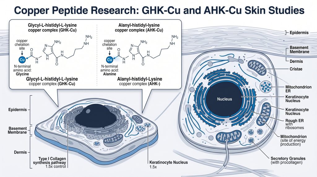
A copper peptide research compound is a small peptide. It chelates a copper(II) ion to form a stable complex. GHK-Cu contains glycine, histidine, and lysine. AHK-Cu replaces glycine with alanine at one position. Both peptides are studied in fibroblast and skin cell models. Lab teams measure collagen output and SOD activity. SOD stands for superoxide dismutase. Research-grade purity above 99% is needed for reliable data.
Next Level Pharm is a US-based supplier of research-grade GHK-Cu, verified to ≥99.4% purity by HPLC and mass spectrometry on every batch. Each lot ships with a COA and a lot number for full traceability.
Key Takeaways
- GHK-Cu and AHK-Cu share copper-binding affinity: Both peptides chelate copper(II) ions. They differ at one N-terminal amino acid.
- SPARC and TGF-beta are key targets: GHK-Cu modulates SPARC and TGF-beta in fibroblast models. AHK-Cu has been studied in keratinocyte cell assays.
- Collagen and SOD are primary biomarkers: Labs measure collagen I and III by ELISA. SOD activity assays track antioxidant response in treated cells.
- COA-verified lots: Next Level Pharm ships lyophilized GHK-Cu lots with HPLC and mass spec data on every batch. Each lot has a traceable number.
- Research use only: All GHK-Cu in this catalog is for laboratory research use only. No clinical outcomes are claimed.
What Are GHK-Cu and AHK-Cu at the Molecular Level?
GHK-Cu is a tripeptide copper complex. AHK-Cu differs at the N-terminal amino acid only.
According to Pickart and Margolina (2018), GHK-Cu contains glycine, histidine, and lysine. The histidine ring coordinates the copper(II) ion. Lysine and glycine stabilize the chelate in solution. AHK-Cu replaces glycine with alanine. This change does not alter copper-binding affinity much. Both complexes carry a charge that aids cell uptake. In cell media, albumin carries free copper ions. Albumin buffers free copper and protects cells. Researchers add defined copper to form the complex. This step ensures stable complex formation before cell exposure.
How Does GHK-Cu Activate SPARC and TGF-Beta Pathways?
GHK-Cu upregulates SPARC and activates TGF-beta. This has been studied in WI-38 fibroblast cell lines.
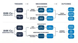
According to Pickart (2008), GHK-Cu interacts with the SPARC protein pathway. SPARC is a protein that regulates collagen assembly. In WI-38 fibroblast cells, GHK-Cu raises SPARC levels. SPARC then promotes TGF-beta1 signaling. TGF-beta1 activates Smad2 and Smad3 proteins. Smad2/3 then drives collagen I and III gene output. Researchers confirm this by ELISA on cell media. IMR-90 cells show a similar collagen response. Fibronectin expression is also tracked by western blot. Fibronectin is an extracellular matrix protein. Western blot uses anti-fibronectin antibodies on cell lysate. Band density is normalized to beta-actin as a loading control.
How Does AHK-Cu Compare With GHK-Cu in Cell Models?
AHK-Cu and GHK-Cu have similar copper affinity. They differ in cell pathway activity across models.
According to Maquart et al. (1999), fibroblast wound models respond to copper peptides. AHK-Cu has been studied in HaCaT keratinocyte cells. HaCaT is a human keratinocyte cell line. AHK-Cu raises fibronectin in HaCaT cultures. GHK-Cu shows stronger TGF-beta effect in fibroblast models. AHK-Cu activity is stronger in keratinocyte assays. Both peptides shift MMP expression at low levels. MMP stands for matrix metalloproteinase. MMP-1 and MMP-2 expression is tracked by qPCR. The table below compares both compounds. Researchers run parallel assays on both cell lines to confirm model-specific differences.
| Feature | GHK-Cu | AHK-Cu |
| Structure | Glycyl-histidyl-lysine + Cu(II) | Alanyl-histidyl-lysine + Cu(II) |
| N-terminal amino acid | Glycine | Alanine |
| Primary cell model | WI-38, IMR-90 fibroblasts | HaCaT keratinocytes |
| Receptor pathway | SPARC, TGF-beta, Smad2/3 | Fibronectin, MMP modulation |
| Primary biomarker | Collagen I/III by ELISA | Fibronectin by western blot |
| SOD activity assay | Yes, reported | Limited published data |
All data are from cell culture research models. No human outcome claims are made.
What Biomarker Assays Are Used in Copper Peptide Research?
Labs use ELISA, western blot, and SOD assays. qPCR tracks MMP expression changes in treated cells.
Collagen I and III are measured by sandwich ELISA. The assay runs on cell media after treatment. Fibronectin is detected by western blot. Anti-fibronectin antibodies are used for detection. SOD activity assays measure antioxidant enzyme response. SOD activity is given in units per mg protein. MMP-1 and MMP-2 gene expression is tracked by qPCR. qPCR needs RNA extraction and reverse transcription first. All assays need a COA-verified copper peptide lot. A lot below 99% purity can shift ELISA and SOD readings. Researchers also run a Bradford protein assay to normalize SOD data. Protein normalization reduces variability between replicate wells.
How Is Copper Transport Managed in Cell Culture Studies?
Albumin and ceruloplasmin carry copper in cell media. They buffer free copper ion levels during studies.
Free copper ions are toxic to cells at high levels. Albumin is the main copper carrier in serum media. It chelates free copper ions. This keeps free-ion levels low and safe for cells. Ceruloplasmin is a serum protein that also binds copper. In serum-free assays, researchers add albumin to control copper. Copper peptides like GHK-Cu compete with albumin for copper. The balance shifts with media pH. Researchers set media pH to 7.4 for stable results. They also confirm complex stability by UV-Vis test.
What Research Tools Are Available for GHK-Cu Studies?
Copper peptide studies need a COA-verified lot. ELISA, SOD assay, and western blot kits are the core tools.
Research teams need a verified GHK-Cu lot to run any study. The lot must match the published reference standard for molecular weight and purity. Next Level Pharm verifies each GHK-Cu lot by HPLC and mass spectrometry before shipping. Collagen I and III ELISA kits for cell media are sold by major suppliers. SOD activity kits use a colorimetric substrate to detect enzyme levels. Anti-fibronectin antibodies for western blot must be validated against a human standard. View the GHK-Cu product page for lot-specific COA and mass spec data.
Frequently Asked Questions
What is GHK-Cu in peptide research?
GHK-Cu is a tripeptide copper complex. It contains glycine, histidine, and lysine. The histidine ring coordinates a copper(II) ion. It is studied in WI-38 and IMR-90 fibroblast cells. Lab teams measure SPARC, TGF-beta, and collagen output. GHK-Cu is for laboratory research purposes only. It is not approved for human use.
How does AHK-Cu differ from GHK-Cu structurally?
AHK-Cu replaces glycine with alanine at the N-terminus. This is a single amino acid change. It does not greatly change copper-binding affinity. The histidine still coordinates the copper(II) ion. AHK-Cu is more often studied in HaCaT cells. GHK-Cu is studied more in fibroblast cell lines. Their downstream signaling profiles differ between these models.
What is SPARC and why does it matter for copper peptide research?
SPARC stands for secreted protein acidic and rich in cysteine. It is also called osteonectin. SPARC regulates collagen fibril assembly. GHK-Cu raises SPARC levels in fibroblast cell models. SPARC then promotes TGF-beta1 signaling. TGF-beta1 activates Smad2/3 proteins. Smad2/3 drives collagen I and III gene output. This cascade is measured in WI-38 cells.
What cell lines are used in copper peptide research?
WI-38 and IMR-90 are common in GHK-Cu studies. WI-38 is a human lung fibroblast cell line. IMR-90 is a human fetal lung fibroblast line. Both produce collagen and fibronectin as biomarkers. HaCaT is an immortalized human keratinocyte cell line. It is used in AHK-Cu studies. Each line allows collagen, fibronectin, and MMP measurement.
How is collagen synthesis measured in GHK-Cu cell studies?
Collagen I and III are measured by sandwich ELISA. The assay runs on media collected after treatment. Anti-collagen antibodies capture secreted collagen. A detection antibody signals the output level. Results are in micrograms of collagen per ml. Western blot also confirms collagen I protein bands. Both methods need a high-purity copper peptide lot. Researchers run a standard curve for each ELISA plate. The curve maps absorbance values to collagen concentration.
What does SOD activity indicate in copper peptide assays?
SOD stands for superoxide dismutase. It is an antioxidant enzyme in cells. SOD converts superoxide radicals to hydrogen peroxide. In GHK-Cu research, SOD assays track antioxidant response. Higher SOD activity suggests an antioxidant enzyme shift. SOD activity is measured in units per mg protein. This is a common readout in published fibroblast studies. Labs run duplicate wells and average the readings for reliability.
Why is copper transport important in cell culture assays?
Free copper ions are toxic to cells at high levels. Albumin and ceruloplasmin buffer free copper in media. Albumin chelates copper ions to keep levels safe. Copper peptides compete with albumin for free copper. Researchers control this by setting media pH to 7.4. They also test complex stability by UV-Vis before adding to cells. This step confirms copper is bound to the peptide. UV-Vis absorbance at 570 nm confirms the complex. A flat baseline shows no free copper ions remain.
Are copper peptides approved for human use?
No. GHK-Cu and AHK-Cu are not approved for human use. They are for laboratory research only. Researchers must follow all applicable guidelines. No health or outcome claims are made here. All lots ship with a COA and lot number. The COA includes HPLC and mass spec data. This is for research traceability only.
Summary
GHK-Cu and AHK-Cu are copper peptide research compounds. They share copper-binding affinity but differ at one amino acid. GHK-Cu activates SPARC and TGF-beta in fibroblast models. AHK-Cu shows fibronectin and MMP activity in keratinocyte cells. No human outcome claims are made here.
Key biomarkers include collagen I and III by ELISA. Fibronectin is tracked by western blot. SOD activity assays confirm antioxidant response. MMP expression is tracked by qPCR. A COA-verified lot at 99% purity is needed. High purity gives reliable, publishable data.
Assay methodology shapes data quality in copper peptide studies. ELISA standard curves must be validated for each plate run. Western blot band density is normalized to beta-actin. SOD activity values are reported per mg of total protein. These controls reduce inter-assay variability across experiments.
What Should You Do Next?
- Review the lot COA for your GHK-Cu or AHK-Cu lot before any cell assay.
- Confirm HPLC purity and mass spec molecular weight against the reference standard.
- Consult published fibroblast and keratinocyte protocols on PubMed before your study.
- Request PhD research support if your lab needs guidance on ELISA or SOD assay setup.
- Browse beauty research peptides for related research tools.
- Shop research peptides at https://nextlevelpharm.com/shop/.
People Also Read
- SNAP-8 Research: SNARE Complex and Skin Cell Studies
- Glutathione Research: Antioxidant Tripeptide Cell Studies
About the Author
Next Level Pharm Research Team
Alex M covers peer-reviewed findings in peptide science for Next Level Pharm, a US-based supplier of research-grade peptides verified to ≥99% purity via HPLC and mass spectrometry on every batch.
Disclaimer: The information provided on this page is for educational and research purposes only. Next Level Pharm products are intended for laboratory research use only. They are not intended for human consumption, diagnostic, therapeutic, or medicinal purposes. This content does not constitute medical advice. Always consult a licensed healthcare professional before making any health-related decisions.