
Peptide Microarray Research: High-Throughput Binding

Last updated: October 2026
A peptide microarray is a solid platform that holds thousands of short peptide sequences at defined spots on one surface. Each sequence sits at a fixed location, and a labeled target is added to find binding partners. This format screens an entire peptide library against one target in a single run.
Next Level Pharm supplies research-grade peptides with a verified purity of 99.4% across 100 recent batches. All vials are tested by HPLC and mass spectrometry. Each order ships with a COA and third-party testing records access.
This format gives labs a fast way to map binding motifs at scale. Many peptide candidates can be compared in one study. The resulting data helps isolate sequences that need deeper testing in cell models.
Key Takeaways
- High-Throughput Parallel Analysis: Peptide arrays let researchers test thousands of binding events on one chip. This saves time over running separate assays for each candidate.
- Surface Immobilization Principle: The method anchors a full peptide library onto a solid base. Specific binding events are then detected with high spatial resolution.
- Diverse Research Uses: Scientists apply these arrays for B-cell epitope mapping and enzyme profiling. They also help identify biomarkers in complex biological samples.
- Specialized Data Processing: Accurate results require software for image reading and noise removal. Statistical tests then confirm each binding signal.
- Flexible Fabrication Methods: Researchers build arrays by spotting pre-made peptides or by growing chains on a surface. Both methods produce dense, uniform screening libraries.
These points give a clear base for reading molecular binding in the lab. The sections below explain how peptide structure affects binding and signal output at each step.
How do peptide microarrays work?
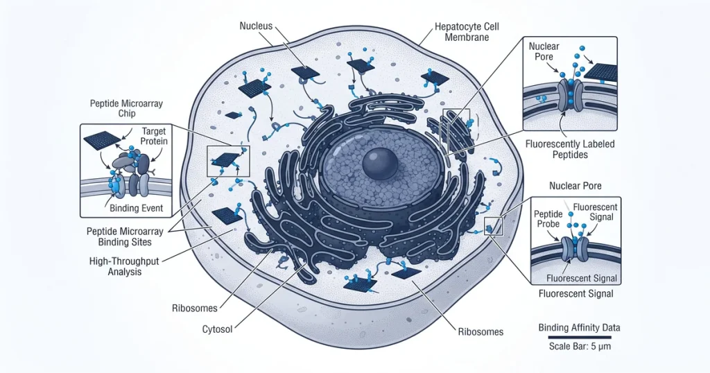
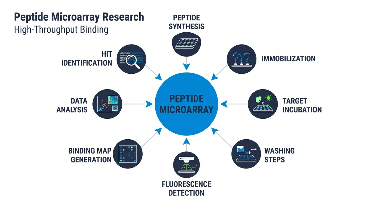
An array works by anchoring thousands of short peptide sequences to a solid chip at defined spots. A labeled target molecule is then passed over the surface. It binds to matching peptides during a set timed step.
After this step, scientists wash away all unbound molecules. Only stable bonds remain on the chip. A laser scanner then reads the signal at each spot.
According to PubMed (2020), this format lets labs screen large libraries with high resolution. Scientists use the resulting binding map to find sequences for further study in isolated cell models.
What types of peptide arrays exist?
Two main types of peptide arrays are used in the lab. Spot methods place pre-made peptides onto a slide at set locations. In situ synthesis builds each chain on the chip, one amino acid at a time.
Spot methods are flexible and work well for small studies. Researchers select from existing stocks and deposit sequences with precision. Spot density is lower than with in situ methods.
In situ synthesis is standard for high-density work. Photolithography uses light and masks to build chains at exact surface sites. A maskless synthesizer provides finer control through digital light patterns.
How are binding events detected?
Binding events are detected by measuring contact between a peptide and a target molecule. Fluorescence detection is the most common lab approach. A dye is attached to the target before it is placed on the chip.
After the timed step, the system reads the light at each spot. Higher signal means stronger binding at that peptide site. This method is fast and suited to large arrays.
Label-free methods avoid adding dye to the target. Surface Plasmon Resonance (SPR) measures small mass changes on a sensor as molecules bind. SPR avoids any signal noise from chemical labels.
Researchers sourcing research peptides for binding studies can browse the full catalog at Next Level Pharm.
What does microarray data analysis involve?
Array data analysis starts with reading signal intensity from scanned chip images. Software calculates the value at each spot on the surface. High values show that the target bonded to the peptide at that site.
Background removal strips noise from non-specific signal across the chip. Spot correction adjusts for uneven signal levels across the slide. According to NCBI (2019), these steps allow accurate comparison across separate experimental runs.
Statistical tests then find hits. Hits are sequences with signal well above the noise threshold. Final data reflects true binding events, not imaging artifacts.
What are key research applications?
Arrays serve several core functions in lab research. B-cell epitope mapping finds the exact sequences that trigger an immune response. Enzyme substrate profiling locates the active sites of target enzymes.
A third use is antibody cross-reactivity screening to confirm binding specificity. According to Molecules (2024), biomarker discovery is a growing use for these platforms. Arrays identify unique peptide patterns in plasma or serum samples.
Key research uses include:
- B-cell epitope mapping for immune response studies.
- Enzyme substrate profiling to locate active binding sites.
- Antibody cross-reactivity screening for specificity tests.
- Biomarker discovery in plasma and serum sample sets.
Next Level Pharm stocks COA-verified research peptides for receptor-binding and related array-based lab studies.
What are the limitations of this method?
The main limits include non-specific binding, altered peptide shape, and synthesis gaps. Non-specific binding occurs when a target attaches to the slide surface rather than a peptide. This creates noise that can hide true binding signals.
Short peptides fixed to a surface may also lose their native shape. This altered shape can limit how well a peptide binds to its target. These shape issues affect data quality in some experiment types.
Array-to-array differences can also affect cross-batch comparisons. Gaps in synthesis fidelity create uneven spot quality between chip batches. Standard protocols reduce these common technical problems.
| Detection Method | Label Required | Sensitivity | Common Application |
|---|---|---|---|
| Fluorescence | Yes | High | Epitope mapping, screening |
| SPR | No | Very high | Kinetics, affinity constants |
| Colorimetric | Yes | Medium | Low-throughput confirmatory tests |
| Chemiluminescence | Yes | High | High dynamic range studies |
| Mass Spectrometry | No | Very high | Label-free identification |
Frequently Asked Questions
What is the principle of peptide microarray technology?
An array holds thousands of short peptide sequences on a dense solid surface. A target sample passes over the surface and binds to matching spots. Sensors measure the strength and location of each binding event.
This parallel format screens many sequences in one run. Researchers get a broad view of how one target binds across an entire library. Single-sequence tests cannot match this throughput.
How are peptides attached to the array surface?
Peptides attach to the surface through covalent bonding or physical adsorption. Covalent links form between groups on the peptide, such as cysteine or lysine, and reactive sites on the slide coating. This bond resists repeated washing steps in the assay.
Spot-on methods deposit pre-made peptides directly onto the surface. Proper alignment keeps the binding region of each peptide exposed. Both methods keep binding motifs accessible to the target.
What binding interactions can peptide microarrays detect?
These arrays detect binding between peptides and antibodies, enzymes, or receptors. Researchers use them to map sites for serum antibodies in disease studies. They also find protein interaction domains across large sequence sets.
The platform tracks enzyme changes, such as phosphorylation, by watching peptide-kinase reactions. It maps thousands of events in a single run. This breadth helps characterize complex biological pathways.
How is microarray signal data analyzed for binding affinity?
Signal analysis begins with reading the intensity at every spot on the chip. Each value is compared to a background control to estimate binding strength. Software aligns the grid and calculates binding density for each spot.
This links signal to known concentration amounts. Researchers use this data to rank peptides by affinity across the full library. High-intensity spots mark the sequences with the strongest binding.
What controls are needed in peptide microarray experiments?
Both negative and positive controls are required in any array experiment. Negative controls are blank spots or peptides with no affinity for the target. They set the background binding level on the chip.
Positive controls are peptides known to bind the target strongly. They confirm the detection system is working as expected. Together, these standards let researchers normalize signals and confirm results.
Can peptide microarrays provide quantitative data?
Yes, these arrays yield quantitative data under controlled conditions. Scientists measure signal rates or use calibration curves to estimate affinity constants. By tracking binding phases, researchers calculate kinetics for each peptide spot.
This approach goes beyond simple yes-or-no detection. Researchers see the speed and strength of each binding event. Quantitative array data can be compared to results from single-peptide assays.
What is the difference between a peptide and a protein microarray?
Protein arrays display full proteins or large domains that keep complex three-dimensional shapes. Peptide arrays feature short, linear sequences that often lack defined structure. The key difference is the size and fold state of each displayed molecule.
Researchers choose peptide arrays to map linear epitopes or short binding motifs. Protein arrays suit studies that require intact folded proteins. Each format serves a distinct research purpose.
How does photolithography create peptide arrays?
Photolithography uses light and masks to build peptides on a chip surface. A light-sensitive group covers the slide and blocks reactions at every site. Light hits specific zones to remove the blocking group at those sites.
The next amino acid binds only at the exposed sites. Repeating this with new masks and amino acids builds distinct peptide chains. This layer-by-layer method creates high-density custom libraries.
How is non-specific binding reduced during array experiments?
Non-specific binding is reduced by applying blocking buffers that coat empty slide areas. These buffers contain bovine serum albumin (BSA) or surfactants. They stop target molecules from sticking to the support rather than the peptides.
Adjusting salt levels or wash buffer pH removes weak, short-lived bonds. Careful washing keeps only high-affinity binding events on the surface. This step is performed before final signal detection.
What are the advantages of in situ synthesis for peptide arrays?
In situ synthesis creates high-density arrays with precise control over each sequence. Chains are built on the slide rather than deposited from solution. Spot density is much higher than in spot-on-demand methods.
This approach cuts the need for large pre-made peptide stocks. Researchers can build custom libraries quickly. Many amino acid variants can be tested in one batch to find binding motifs.
Summary
Peptide microarray binding research gives scientists a scalable method for studying molecular binding in the lab. By anchoring thousands of short sequences on one surface, labs can screen many peptides against a target in one run. Sensors map binding events, and data analysis confirms true hits.
Key uses include B-cell epitope mapping, enzyme substrate work, and biomarker discovery. COA-verified, HPLC-tested research peptides are available for these and related array-based research programs.
What Should You Do Next?
Researchers planning array studies should confirm that selected peptides match the specific model and target in use. Checking the COA for each batch confirms that every vial meets the required purity standard. Keeping written handling protocols helps maintain material integrity across the study.
Next Level Pharm provides research-grade peptides with full COA and third-party testing records access for every order. Researchers can shop the full peptide catalog to find the right compound for their specific assay.
People Also Read
- Circular Dichroism in Peptide Research: CD Spectroscopy Methods
- MAPK/ERK Pathway Peptide Research: Kinase Cascade Analysis
About the Author
Next Level Pharm Research Team
The Next Level Pharm research team is composed of biochemists and laboratory scientists dedicated to providing researchers with the highest-purity, lab-verified research peptides available. Every batch is HPLC and mass spec confirmed before dispatch.
Disclaimer: The information provided on this page is for educational and research purposes only. Next Level Pharm’s products are intended for laboratory research use only. They are not intended for human consumption, diagnostic, therapeutic, or medicinal purposes. This content does not constitute medical advice. Always consult a licensed healthcare professional before making any health-related decisions.