
What Are b and y Ions in Peptide Mass Spectrometry?

Last updated: July 2026
Ion fragmentation in mass spectrometry is a method used to confirm the amino acid sequence of a research peptide. A peptide is broken into smaller charged pieces called fragment ions. The pattern of these ions matches the amino acid sequence. According to Roepstorff P & Fohlman J (1984) (PMID 6525415), the b-ion and y-ion system is the standard for labeling peptide fragment ions. This system has been used for over four decades.
Next Level Pharm is a US-based supplier of research-grade peptides. Every lot is confirmed by HPLC purity and mass spectrometry identity testing. A COA with a lot of ships with every order.
Understanding ion fragmentation helps researchers read mass spec COA data. It also helps them evaluate the identity method used by a peptide supplier.
Key Takeaways
- b Ions Come From the N-Terminal End: A b ion contains the N-terminal (beginning) portion of the peptide. It forms when a peptide bond breaks and the charge stays on the N-terminal fragment.
- y Ions Come From the C-Terminal End: A y ion contains the C-terminal (end) portion of the peptide. It forms when the charge stays on the C-terminal fragment after bond cleavage.
- Tandem MS Creates the Fragment Spectrum: Tandem mass spectrometry (MS/MS) selects a parent ion. It then fragments that ion. The result is a spectrum of smaller ions called fragment ions.
- CID Is the Standard Fragmentation Method: CID (collision-induced dissociation, a method where ions collide with inert gas to fragment) produces b and y ions. It is the most common method in peptide quality testing.
- Fragment Patterns Confirm Amino Acid Sequence: Each peptide has a unique b-ion and y-ion pattern. Matching this pattern to the expected sequence confirms peptide identity at the sequence level.
Ion fragmentation confirms more than molecular weight. It confirms the exact amino acid order. This rules out deletion sequences and truncation artifacts from synthesis.
What Are b Ions and y Ions?
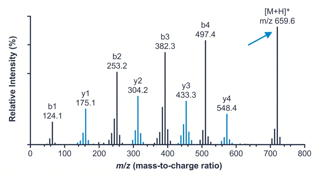
A b ion includes amino acid residues from the N-terminal end of the peptide. A y ion includes residues from the C-terminal end. The mass difference between two adjacent b ions equals the mass of one amino acid residue. Researchers use this to read the sequence from the spectrum. Together, both ion series can cover the full peptide sequence.
Each amide bond in the peptide can break during fragmentation. When the bond between residue 3 and residue 4 breaks, a b3 ion and a y(n-3) ion form. Here, n is the total residue count. According to Steen H & Mann M (2004) (PMID 15340377), b and y ions are the primary tools for reading a peptide sequence. A tandem mass spectrum provides this data. Other ion types (a, c, x, z) exist but are less common in standard CID experiments.
How Does Tandem Mass Spectrometry Identify a Peptide?
Tandem mass spectrometry uses two stages of mass analysis. In the first stage, one precursor ion is selected by its mass-to-charge ratio (m/z). In the second stage, the precursor is fragmented. The fragment ions are measured and plotted as a spectrum. This spectrum is matched to the expected pattern for the target sequence.
The first stage acts as a filter. It isolates one ion from the sample. In COA testing, this is the intact peptide. The second stage breaks it apart. The spectrum is then compared to calculated masses. A good match confirms both molecular weight and amino acid sequence. MS/MS is stronger than MS1-only testing.
What Is the Fragmentation Pattern?
The fragmentation pattern is the set of b-ion and y-ion peaks in an MS/MS spectrum. Each peptide sequence gives a unique set of fragment masses. This pattern acts as a fingerprint. Comparing the measured pattern to the expected pattern confirms whether the peptide has the right sequence.
For a 10-residue peptide, there are 9 possible b ions and 9 possible y ions. Not all appear in every spectrum. Some bonds fragment more easily than others. Proline residues increase fragmentation at the bond on their N-terminal side. This gives strong y ions at proline positions. Researchers use these trends to interpret spectra where some expected peaks are absent.
How Does CID Generate Fragment Ions?
CID (collision-induced dissociation) accelerates the precursor ion into an inert gas. The gas is usually argon or nitrogen. The energy breaks the amide bonds in the peptide chain. This produces b and y ions. The collision energy is adjusted for each peptide mass. Too little energy leaves the peptide intact. Too much energy breaks fragments into very small ions.
According to Mischak H et al. (2012) (doi:10.1016/j.jprot.2012.01.012), CID is the most used fragmentation method in quantitative peptide research. It is compatible with most mass spectrometer platforms. Instruments use normalized collision energy settings to optimize fragment yield across different peptide masses.
How Do Fragment Ions Confirm Peptide Identity?
Fragment ions confirm identity by matching measured masses to calculated masses. For each b and y ion, a calculated m/z value exists based on the amino acid residue masses. If the measured ions match the calculated m/z values within 0.02 Da, the identity is confirmed. This is the standard for high-resolution instruments.
High-resolution mass spectrometers achieve accuracy below 5 parts per million (ppm). This level of precision rules out most false sequence matches. For research peptide quality testing, a confirmed identity result needs two things. It needs a match on the intact mass (MS1) and at least 5 to 8 fragment ions (MS2). A score is computed based on how many expected ions were found and how well they match.
How Do Suppliers Use Ion Fragmentation in QC Testing?
Peptide suppliers use MS/MS ion fragmentation to confirm the sequence of each synthesis batch. The batch is run by LC-MS/MS. The fragmentation spectrum is matched to the expected b and y ions for that sequence. This confirms the amino acid order, not just the molecular weight. It rules out deletion sequences. Deletion sequences can have the same molecular weight but a different sequence.
HPLC purity and mass spec identity testing are separate tests. HPLC checks the target compound proportion in the sample. Mass spectrometry confirms the amino acid sequence. A COA with MS/MS fragment ion data is stronger than one that only reports an intact molecular weight. Shop research peptides at Next Level Pharm. COA documentation with HPLC purity, mass spectrometry identity data, and lot number ships with every batch.
| Ion Type | Origin End | Contains | Common in CID? |
| b ion | N-terminus | N-terminal residues | Yes |
| y ion | C-terminus | C-terminal residues and H2O | Yes |
| a ion | N-terminus | b ion minus CO (28 Da) | Sometimes |
| z ion | C-terminus | y ion minus NH3 (17 Da) | Less common |
| c ion | N-terminus | b ion plus NH3 | ETD only |
| x ion | C-terminus | y ion plus CO2 minus H | Rare |
Frequently Asked Questions
What Is a Precursor Ion?
A precursor ion is the intact peptide ion selected for fragmentation in the first stage of tandem mass spectrometry. It is selected by its mass-to-charge ratio (m/z). In peptide COA testing, the precursor is the full-length target peptide. Once selected, it is fragmented by CID or another method. The precursor mass from MS1 data confirms the molecular weight. The fragment ions from MS2 data confirm the amino acid sequence.
What Does m/z Mean in Mass Spectrometry?
m/z stands for mass-to-charge ratio. It is the core measurement in mass spectrometry. A mass spectrometer detects ions based on their mass divided by their charge. A peptide with a mass of 1000 Da and a charge of plus 2 appears at m/z 500.5 (accounting for proton mass). Charge states range from plus 1 to plus 4 or higher for larger peptides. Multiple charge states for the same peptide appear as a series of peaks in the MS1 spectrum.
What Is a Deletion Sequence?
A deletion sequence is a peptide that is missing one amino acid residue from the target sequence. It forms when one coupling step fails during solid-phase synthesis. Deletion sequences can have the same or similar molecular weight as the target. HPLC may not separate them if their retention times are close. MS/MS fragment ion data detects deletion sequences because their fragment ion pattern shifts at the missing residue position. This is why MS/MS data is preferred over MS1-only identity testing.
Why Are Some Fragment Ions Missing From the Spectrum?
Some amide bonds fragment more easily than others. Bonds near proline, aspartic acid, or glutamic acid tend to break readily. These give strong fragment ions. Bonds in the middle of hydrophobic regions may fragment less. Internal fragment ions can also appear and complicate the spectrum. For short peptides under 10 residues, most expected ions are visible. For longer peptides, partial ion coverage is normal. Identity confirmation does not require all ions to be present.
What Is the Difference Between MS1 and MS2 Data?
MS1 is the first mass spectrum. It shows the intact peptide ions in the sample and gives the molecular weight. MS2 (also called MS/MS or tandem MS) is the second spectrum. It shows fragment ions from a selected precursor and gives the amino acid sequence. A COA with only MS1 data confirms molecular weight but not sequence. A COA with both MS1 and MS2 data confirms both. MS2 data is the stronger identity test for research peptides.
What Is Mass Accuracy?
Mass accuracy is how close the measured m/z of an ion is to the calculated m/z. It is expressed in parts per million (ppm) or millimass units (mDa). High-resolution instruments (Q-TOF or Orbitrap) achieve below 5 ppm accuracy. At 5 ppm for a 1000 Da ion, the mass window is 0.005 Da. This narrow window eliminates most false sequence matches. Low-resolution instruments achieve 0.1 to 0.5 Da accuracy. This is less reliable for sequence confirmation against similar compounds.
What Is a Sequence Ladder?
A sequence ladder is the complete series of b ions or y ions in an MS/MS spectrum. Reading the b ion series from smallest to largest (b2, b3, b4…) gives the N-terminal sequence. Reading the y ion series gives the C-terminal sequence. A full ladder covers every residue position. Most spectra have partial ladders. A partial ladder covering 60 to 70 percent of positions is enough for high-confidence sequence confirmation in research peptide quality testing.
Can Mass Spectrometry Detect Amino Acid Modifications?
Yes. Modifications change the mass of a specific residue. Common modifications in research peptides include oxidation of methionine (plus 16 Da) and acetylation (plus 42 Da). If a modification is present, the b or y ion masses shift at the modified residue position. This shift localizes the modification to a specific amino acid. MS1 alone can detect that a modification is present but cannot identify which residue is modified. MS/MS provides residue-level localization.
What Fragment Ion Data Should Appear on a COA?
A COA with full MS/MS data should include the precursor ion m/z and charge state, a list of major fragment ions detected, the measured vs. calculated mass for each ion, and a sequence coverage percentage. Some COAs include a spectrum image. A confirmed sequence match is usually stated as sequence confirmed by LC-MS/MS. A COA with only an MW confirmed result without MS/MS data does not include sequence-level confirmation for the research peptide lot.
Summary
Ion fragmentation in mass spectrometry breaks a peptide into b ions and y ions. The b ions contain the N-terminal end. The y ions contain the C-terminal end. The pattern of these ions is unique to the amino acid sequence.
CID is the standard fragmentation method. It collides the precursor ion with an inert gas to break amide bonds. A confirmed identity result needs at least 5 to 8 fragment ions to match the target sequence. This confirms sequence identity, not just molecular weight.
What Should You Do Next?
When reviewing a peptide COA, check whether mass spectrometry data includes MS/MS fragment ions or only an intact molecular weight (MS1) result. MS/MS data confirms the amino acid sequence. MS1 alone confirms only the molecular weight. For research applications where sequence accuracy matters, request MS/MS data or the fragment ion list from the supplier.
Shop research peptides. Every batch at Next Level Pharm includes HPLC purity, mass spectrometry identity data, and lot number on the COA.
People Also Read
- What Is Mass Spectrometry? How It Confirms Peptide Identity
- Peptide Purity Testing: What HPLC and Mass Spec Confirm
About the Author
Next Level Pharm Research Team
Alex M covers peer-reviewed findings in peptide science for Next Level Pharm, a US-based supplier of research-grade peptides verified to ≥99% purity via HPLC and mass spectrometry on every batch.
Disclaimer: The information provided on this page is for educational and research purposes only. Next Level Pharm products are intended for laboratory research use only. They are not intended for human consumption, diagnostic, therapeutic, or medicinal purposes. This content does not constitute medical advice. Always consult a licensed healthcare professional before making any health-related decisions.