
GLP-1 Receptor Structure: Class B GPCR Research Explained

GLP-1 Receptor Structure: Class B GPCR Research Explained
Last updated: September 2026
A GLP-1 receptor structure is that of a Class B G-protein coupled receptor, a family of proteins that span the cell membrane and convert extracellular signals into intracellular responses. This set protein architecture plays a primary role in how hormonal ligands interact with cell pathways, managing whole-body signals. Current research finds this receptor as a critical component in the study of energy control and glucose homeostasis within controlled lab settings with 70+ active peptide SKUs now ready. Researchers have access to a wide array of compounds for their research designs.
Next Level Pharm gives research-grade peptides with a commitment to test transparency and batch consistency. Each vial is produced to meet a high standard, maintaining a 99.4% average purity level across the last 100 batches. Each order includes a checked certificate of test. Researchers may access lot-set records through a simple online lookup portal at any time.
These standards ensure that each compound remains stable during transit. All products are lyophilized and sealed under an inert atmosphere. Because the peptides are freeze-dried, they remain stable at room temperature, do not need cold chain logistics for successful transport. All inventory is stored within the USA for an approximate 48-hour average dispatch time to meet project timelines.
Key Takeaways
- Receptor Classification: The GLP-1 receptor is a Class B G-protein coupled receptor, which uses a large domain for hormone binding.
- Two-Step Binding: GLP-1 peptides first dock at the extracellular domain before inserting into the transmembrane core for full action.
- High Resolution Visualization: Advanced methods like cryo-electron microscopy help map the structure of the receptor for precise study at the atomic level.
- Rational Design possible: staff use core data to create new agonists with set binding affinities and targeted cell signaling preferences.
- Signaling Pathway Action: Action of the receptor mainly starts the Gs protein pathway, which triggers cyclic AMP output inside target cells.
The following sections test the core contacts and signaling dynamics that characterize GLP-1 receptor research. Applying these findings to set lab plans needs a thorough review of peptide binding properties and batch purity reports.
What defines the GLP-1 receptor’s structure?
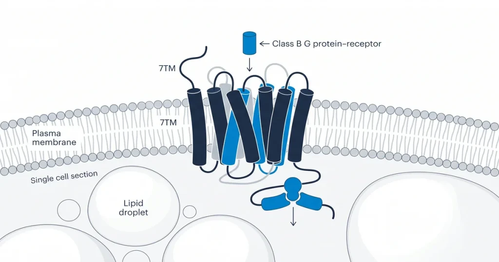
The GLP-1 receptor is defined as a Class B G-protein coupled receptor, which uses a set two-part structure to start cell signaling. This receptor consists of a large N-terminal extracellular domain (ECD) and a seven-transmembrane helix (7TM) domain embedded within the cell membrane. These two regions work in tandem to bind peptide hormones, according to PubMed (2018).
The ECD functions as the initial docking site for the peptide hormone. Once the peptide binds to the ECD, the entire structure goes through a conformational change to trigger intracellular signaling, as noted by PubMed (2018). This mechanism is a hallmark of the Class B receptor family. Semaglutide is ready as a COA-checked research peptide. Each lot is checked by HPLC and mass spec. This 7TM domain is central to how the signal moves from the outside of the cell into the interior.
How does GLP-1 bind to its receptor?
GLP-1 binds to its receptor through a two-step model that ensures precise action of the signaling pathway. Initially, the C-terminal end of the peptide engages with the extracellular domain of the receptor. This first contact is marked by low strength. It serves to orient the peptide correctly for the next phase of binding. Once the C-terminal end is attached, the N-terminal end of the GLP-1 peptide inserts itself into the transmembrane domain pocket. This insertion is the key step, which triggers receptor action and starts the downstream signaling cascade.
Semaglutide is ready as a COA-checked research peptide. Each lot is checked by HPLC and mass spec. This secondary binding event is critical. It forces a conformational change in the receptor structure. Research suggests this movement allows the receptor to interact with intracellular G-proteins, according to PubMed (2018). The spatial arrangement of the transmembrane domain pocket gives a set site for the N-terminal region. Without this set fit, receptor signaling would not occur as intended in test models, maintaining the core health of both the C-terminal and N-terminal ends remains key for accurate peptide-receptor contact studies.
What are Class B GPCRs?
Class B GPCRs are a group of cell surface proteins, binding large peptide hormones like glucagon, GLP-1, and GIP. These receptors are also known as the secretin family of receptors. They function as critical signal transducers within the cell membrane. Staff study these proteins to understand how set ligands interact with cell targets, triggering internal signaling cascades.
These proteins differ from the more common Class A GPCRs in both their structure and binding mechanisms. The defining feature of this group is a large, conserved N-terminal extracellular domain. This domain acts as the primary site for ligand recognition and binding specificity. Because the ligands are large peptide hormones, the receptor needs this unique core surface to ensure proper docking. Once the ligand binds, the receptor changes shape to pass signaling into the cell interior. Tirzepatide is ready as a COA-checked research peptide. Each lot is checked by HPLC and mass spec. The complex interplay between these domains allows for precise control of hormonal responses in various models, according to PubMed (2018).
How is GLP-1R structure research conducted?
Modern research on the GLP-1 receptor structure relies on biophysical techniques like cryo-electron microscopy (cryo-EM) and X-ray crystallography. These tools allow staff to see the receptor at an atomic level, defining how set peptide ligands trigger signaling. By analyzing these complex structures, researchers gain a blueprint for understanding how ligands occupy the transmembrane pocket.
Retatrutide and Tirzepatide are each ready as COA-checked research peptides. Each lot is checked by HPLC and mass spec.
Cryo-EM has become vital for modern study. It allows for the capture of high-resolution snapshots of the receptor. This imaging shows the protein in its various functional states, such as bound to agonists or allosteric modulators. These snapshots reveal how the receptor shifts its shape to convey signals across the cell membrane. Mapping these states helps researchers model how peptide variants interact with the receptor binding site. This lab approach gives the data needed to check how core changes affect ligand binding strength, and downstream cellular responses.
Each lot includes a certificate of test checked by in-house HPLC and mass spectrometry.
Why is the receptor’s structure important for research?
The GLP-1 receptor structure is vital for research. It dictates how peptide agonists interact at the atomic level. A detailed map of the receptor surface is key for the rational design of new compounds. Researchers use these structures to predict how changes to a ligand affect its binding strength to the target. This test helps find the duration of action for a peptide in test models. Understanding these physical shapes is fundamental to modern receptor study and lab study design.
Core maps allow staff to affect the signaling pathway preference of a compound. This focus on biased agonism explores how a ligand can trigger set cell responses, avoiding others. By modifying the peptide chain, researchers can test how certain parts of the receptor affect the outcomes found in a cell test, as noted by NCBI (2020). Tirzepatide is ready as a COA-checked research peptide. Each lot is checked by HPLC and mass spec.
What are allosteric modulators of the GLP-1 receptor?
Allosteric modulators of the GLP-1 receptor are compounds, binding to the receptor at a site distinct from the orthosteric site. The native GLP-1 peptide docks. These compounds affect the action of the receptor without directly competing with the primary ligand. By binding to these alternative zones, allosteric modulators change the shape of the receptor to alter how it responds to normal GLP-1. This indirect affect allows for the control of signaling pathways rather than a simple on or off response.
Positive allosteric modulators (PAMs) serve as a common example. These compounds boost the cell signaling response when the native GLP-1 peptide is present. Research into these small compounds represents a novel approach to fine-tuning receptor function for various investigational studies. Staff explore these contacts to see if receptor response can be adjusted without requiring higher amounts of the primary peptide ligand.
Tirzepatide, Semaglutide, and Retatrutide are each ready as COA-checked research peptides. Each lot is checked by HPLC and mass spec. The study of allosteric sites gives a path to understanding how receptor topography impacts drug efficacy, according to PubMed (2020). By using small compounds to target these regions, researchers can study new methods for selective signaling action in cell-based tests. This area of inquiry continues to grow as new atomic targets are mapped for possible lab application.
Researchers studying GLP-1 receptor mechanisms may also find eloralintide relevant — it is a research-grade GIP/GLP-1 co-agonist analog available as a COA-verified, lyophilized research peptide.
Frequently Asked Questions
What type of receptor is the GLP-1 receptor?
The GLP-1 receptor is a member of the G protein-coupled receptor family, classified as a Class B secretin-like receptor. These proteins are surface receptors that span the cell membrane seven times. They function as sensors for hormones and peptides that float in the extracellular space. Research shows that the GLP-1 receptor binds to glucagon-like peptide-1 and controls signaling pathways, according to PubMed (2018).
How does the GLP-1 receptor signal inside a cell?
When a peptide binds to the receptor, it triggers a change in shape, which activates intracellular G proteins. This action causes an increase in cyclic adenosine monophosphate levels within the cell. This secondary messenger compound then triggers various downstream pathways, affecting enzyme action and gene output. These processes collectively allow the cell to respond to extracellular signals, as cited by NCBI (2020).
What is a Class B GPCR?
A Class B G protein-coupled receptor is a subset of membrane proteins marked by a large extracellular domain. This domain is key for binding large peptide hormones. Unlike other classes of GPCRs, class B receptors often show high specificity for their normal ligands. Scientific literature notes that these receptors are central to endocrine system control, according to the Journal of Peptide Science (2015).
Where are GLP-1 receptors located in the body?
GLP-1 receptors have been detected in many tissues throughout the body, reflecting their diverse control roles. High densities are found in pancreatic beta cells. They affect hormone release. They are also present in the brain, stomach, gut, and heart. Research suggests that this widespread spread allows GLP-1 signaling to affect energy balance across multiple organ systems, as noted by PubMed (2021).
What is the difference between an agonist and an antagonist at this receptor?
An agonist binds to the receptor and triggers a cell response, while mimicking the normal hormone. In contrast, a blocker binds to the receptor but does not activate it. Instead, the blocker blocks the receptor site to prevent other compounds from binding. Studies use both types of compounds to map receptor function and clarify signaling pathways in lab models, according to PubMed (2017).
How do peptides like tirzepatide interact with the GLP-1 receptor?
Tirzepatide acts as a dual agonist by binding to both the GLP-1 receptor and the GIP receptor. This dual engagement creates a distinct signaling profile compared to single-receptor agonists. The compound interacts with the binding pockets of both receptors to start intracellular cascades. Research shows that this combined action affects energy signaling in test models, as shown by PubMed (2021).
What role does the GLP-1 receptor play in insulin secretion research?
The GLP-1 receptor is critical for glucose-dependent insulin release. When the receptor is activated in pancreatic beta cells, it helps to increase the amount of insulin released in response to high glucose levels. This mechanism is a primary focus for researchers studying energy control and signaling pathways. Data from cell tests suggest that this pathway is highly sensitive to outside signals, as cited by PubMed (2019).
Can the GLP-1 receptor be desensitized in lab models?
Yes, the GLP-1 receptor can become reduced through a process called internalization. After prolonged contact to an agonist, the cell removes the receptors from its surface to reduce further signaling. This is a common feedback mechanism that limits overstimulation of the cell. Research models often see this effect, monitoring the duration of receptor action during long-term test trials, according to PubMed (2014).
What is meant by biased agonism at the GLP-1 receptor?
Biased agonism occurs when a ligand selectively activates one signaling pathway over others linked to the same receptor. Instead of triggering a full response, the biased compound favors set downstream signaling. Researchers study this phenomenon to better understand how to refine receptor control in lab tests. This approach gives insight into the potential for selective signaling outcomes within test setups, according to Molecules (2022).
What Should You Do Next?
Review your lab plans to test. All research factors align with your current test goals. Compare your compound’s purity data against the certificate of test provided with your shipment, ensuring it meets your accuracy standards for atomic study. Cross-reference your stored batch lot number with the ready online records, keeping full transparency about the lab makeup of your items.
Researchers sourcing energy peptides can browse the catalog at Shop. Each vial provided by Next Level Pharm ships with a checked certificate of test. Full lot tracking for your records.
People Also Read
- HGH Fragment 176-191 Research: What Lab model Studies Show
- Sermorelin vs CJC-1295: What Research Shows About Each GHRH variant
About the Author
Author Block
Next Level Pharm Research Team
The Next Level Pharm research team is composed of biochemists and lab scientists dedicated to providing researchers with the highest-purity, COA-verified research peptides available. Every batch is HPLC and mass spec verified before dispatch.
Disclaimer: The information provided on this page is for educational and research purposes only. Next Level Pharm products are intended for lab research use only. They are not intended for human consumption, diagnostic, therapeutic, or medicinal purposes. This content does not constitute medical advice. Always consult a licensed healthcare professional before making any health-related decisions.